

Characterization and Genomic Analyses of dsDNA Vibriophage vB_VpaM_XM1, Representing a New Viral Family
- PMID: 39330310
- PMCID: PMC11432961
- DOI: 10.3390/md22090429
Characterization and Genomic Analyses of dsDNA Vibriophage vB_VpaM_XM1, Representing a New Viral Family
Abstract
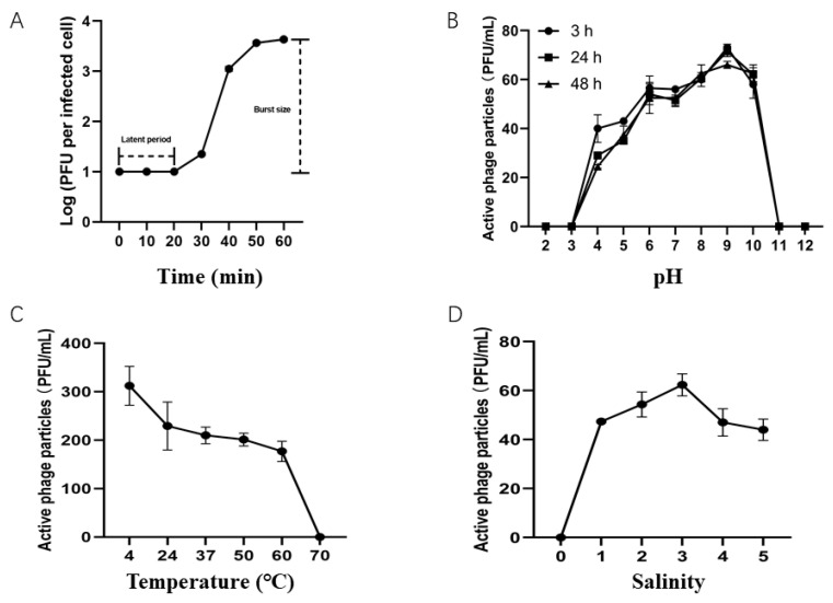
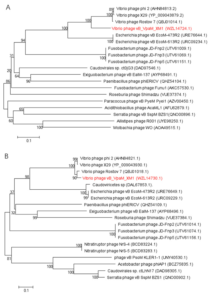
A novel vibriophage vB_VpaM_XM1 (XM1) was described in the present study. Morphological analysis revealed that phage XM1 had Myovirus morphology, with an oblate icosahedral head and a long contractile tail. The genome size of XM1 is 46,056 bp, with a G + C content of 42.51%, encoding 69 open reading frames (ORFs). Moreover, XM1 showed a narrow host range, only lysing Vibrio xuii LMG 21346 (T) JL2919, Vibrio parahaemolyticus 1.1997, and V. parahaemolyticus MCCC 1H00029 among the tested bacteria. One-step growth curves showed that XM1 has a 20-min latent period and a burst size of 398 plaque-forming units (PFU)/cell. In addition, XM1 exhibited broad pH, thermal, and salinity stability, as well as strong lytic activity, even at a multiplicity of infection (MOI) of 0.001. Multiple genome comparisons and phylogenetic analyses showed that phage XM1 is grouped in a clade with three other phages, including Vibrio phages Rostov 7, X29, and phi 2, and is distinct from all known viral families that have ratified by the standard genomic analysis of the International Committee on Taxonomy of Viruses (ICTV). Therefore, the above four phages might represent a new viral family, tentatively named Weiviridae. The broad physiological adaptability of phage XM1 and its high lytic activity and host specificity indicated that this novel phage is a good candidate for being used as a therapeutic bioagent against infections caused by certain V. parahaemolyticus strains.
Keywords: Vibrio parahaemolyticus; bacteriophage; genome analysis; new viral family.
Conflict of interest statement
The authors declare no conflicts of interest.
Figures


Similar articles
-
Isolation and characterization of a novel lytic bacteriophage Pv27 with biocontrol potential against Vibrio parahaemolyticus infections in shrimp.PeerJ. 2025 May 6;13:e19421. doi: 10.7717/peerj.19421. eCollection 2025. PeerJ. 2025. PMID: 40352283 Free PMC article.
-
Characterization and Genomic Analysis of ssDNA Vibriophage vB_VpaM_PG19 within Microviridae, Representing a Novel Viral Genus.Microbiol Spectr. 2022 Aug 31;10(4):e0058522. doi: 10.1128/spectrum.00585-22. Epub 2022 Jul 6. Microbiol Spectr. 2022. PMID: 35862991 Free PMC article.
-
Characterization and genomic analysis of a jumbo phage, PG216, with broad lytic activity against several Vibrio species.Arch Virol. 2025 Jan 6;170(2):31. doi: 10.1007/s00705-024-06215-z. Arch Virol. 2025. PMID: 39762632
-
Genome characterization of novel lytic Myoviridae bacteriophage ϕVP-1 enhances its applicability against MDR-biofilm-forming Vibrio parahaemolyticus.Arch Virol. 2020 Feb;165(2):387-396. doi: 10.1007/s00705-019-04493-6. Epub 2019 Dec 21. Arch Virol. 2020. PMID: 31865470
-
Characterization and genome analysis of lytic Vibrio phage VPK8 with potential in lysing Vibrio parahaemolyticus isolates from clinical and seafood sources.Virol J. 2025 Jan 30;22(1):21. doi: 10.1186/s12985-025-02637-6. Virol J. 2025. PMID: 39885536 Free PMC article.
References
-
- Jeong H.W., Kim J.A., Jeon S.J., Choi S.S., Kim M.K., Yi H.J., Cho S.J., Kim I.Y., Chon J.W., Kim D.H., et al. Prevalence, Antibiotic-Resistance, and Virulence Characteristics of Vibrio parahaemolyticus in Restaurant Fish Tanks in Seoul, South Korea. Foodborne Pathog. Dis. 2020;17:209–214. doi: 10.1089/fpd.2019.2691. - DOI - PubMed
-
- McLaughlin J.B., DePaola A., Bopp C.A., Martinek K.A., Napolilli N.P., Allison C.G., Murray S.L., Thompson E.C., Bird M.M., Middaugh J.P. Outbreak of Vibrio parahaemolyticus gastroenteritis associated with Alaskan oysters. New Engl. J. Med. 2005;353:1463–1470. doi: 10.1056/NEJMoa051594. - DOI - PubMed
MeSH terms
Substances
Grants and funding
LinkOut - more resources
Full Text Sources
Molecular Biology Databases
