. 2024 Sep 7;12(9):655.
doi: 10.3390/toxics12090655.

Integrated Biomarker Response Emphasizing Neuronal Oxidative Stress and Genotoxicity Induced by Oxamyl in Sprague Dawley Rats: Ameliorative Effect of Ginseng as a Neuroprotective Agent

Affiliations

Integrated Biomarker Response Emphasizing Neuronal Oxidative Stress and Genotoxicity Induced by Oxamyl in Sprague Dawley Rats: Ameliorative Effect of Ginseng as a Neuroprotective Agent

Salwa M Abdallah et al. Toxics. .

Abstract

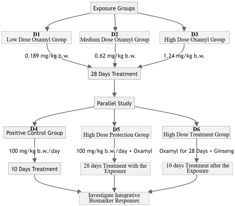
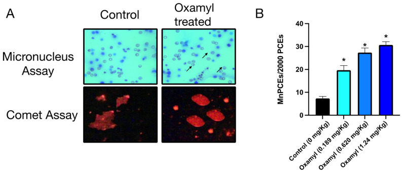
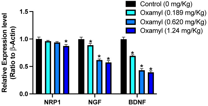
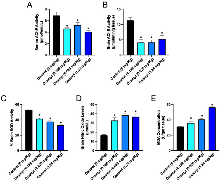
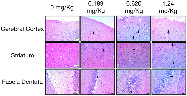
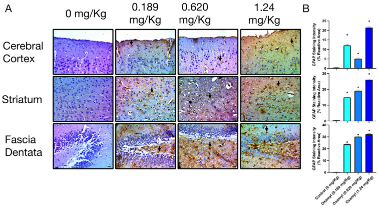
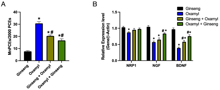
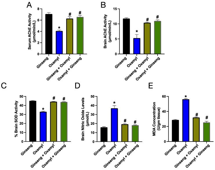
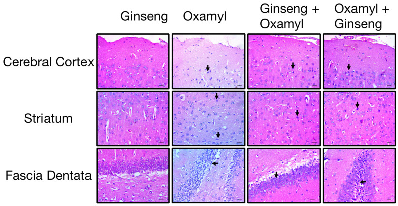
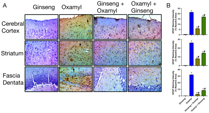
Climate change has led to increased and varying pest infestation patterns, triggering a rise in pesticide usage and exposure. The effects of oxamyl, a widely used nematicide in Egypt, encompasses typical signs of carbamate intoxication; nevertheless, long-term effects of oxamyl exposure, particularly on the nervous system, require further elucidation. This study systematically investigated the mechanism and manifestations of repeated subacute exposure to sublethal doses of oxamyl in male SD rats. Data showed a dose-dependent genotoxic effect, manifested as increased bone marrow micronuclei and decreased brain expression of key genes involved in neurogenesis and neuronal development. Coincidently, brain histopathology showed dose-dependent neurodegeneration in various regions, associated with a significant increase in GFAP immunoreactivity, indicative of neuroinflammation. Biochemical examination revealed a typical pattern of cholinesterase inhibition by carbamates in serum and brain tissue, as well as increased oxidative stress markers in the brain such as SOD activity reduction, alongside an increase in NO and MDA. The ability of Ginseng at a 100 mg/Kg dose to ameliorate the effects of oxamyl exposure was investigated. Ginseng use, either as a protective or therapeutic regimen, attenuated the observed genotoxic, neuroinflammatory, and biochemical alterations. Our results indicate that repeated exposure to oxamyl triggers an integrative neurotoxic response, driven by genotoxicity, oxidative stress, and neuroinflammation, that could trigger an increase in neurological and cognitive disorders. These findings emphasize the urgent need for confirmatory translational studies in human subjects to assess these changes and inform policy decisions regarding safe levels of usage and appropriate agricultural and public health practices.

Keywords: Ginseng extract; Sprague Dawley rats; carbamate pesticide; genotoxicity; integrated biomarker response; neuronal effects; neuroprotection; oxamyl; oxidative stress.


Conflict of interest statement

The authors declare no conflicts of interest.

Figures

Figure 1
Figure 1
Schematic diagram of experimental design.
Figure 2
Figure 2
Results of examination of blood smears from control rats and those treated with oxamyl in the micronucleus test and DNA damage in brain tissue in the comet assay. (A) Representative micrographs showing normal polychromatic erythrocytes in control rats and MnPCEs (arrows) in oxamyl-treated rats (top) and DNA damage staining in the comet assay on brain homogenates from control and oxamy-treated rats (bottom). (B) Summary data of the percentage of MnPECs found in 2000 polychromatic erythrocytes examined in fifteen slides from five different animals in each group. Results are presented as mean ± SEM. Statistical significance was determined using one-way ANOVA followed by Tukey’s multiple comparisons test. p < 0.05 vs. the control is denoted on the graph by *.
Figure 3
Figure 3
Brain expression levels of select genes with a role in neurogenesis in control rats and rats under repeated exposure to different dosing levels of oxamyl. Data are presented as mean ± SEM observations from three different rats per group. Statistical significance was determined using one-way ANOVA followed by Tukey’s multiple comparisons test. * denotes p < 0.05 vs. values from control rats.
Figure 4
Figure 4
Cholinesterase activity in serum (A) and brain tissue (B) as well as markers of oxidative stress including SOD activity (C), nitric oxide levels (D), and MDA levels (E) in brain tissue from control and oxamyl exposed rats. Data are presented as mean ± SEM observations from five different rats per group. Statistical significance was determined using one-way ANOVA followed by Tukey’s multiple comparisons test. * denotes p < 0.05 vs. values from control rats.
Figure 5
Figure 5
Histopathological examination of different brain areas including cerebral cortex (top), striatum (middle), and fascia dentata (bottom) from control rats and rats exposed to different doses of oxamyl. Necrotic neurons and pyknotic nuclei are indicated by arrows. The data shown are representative micrographs from the examination of twelve slides of tissues of three animals per group. Scale bars are 25 μm.
Figure 6
Figure 6
Immunohistochemical examination of GFAP expression in different brain areas including cerebral cortex (top), striatum (middle), and fascia dentata (bottom) from control rats and rats exposed to different doses of oxamyl. (A) Examples of GFAP-positive astrocytes are indicated by arrows. The data shown are representative micrographs from the examination of twelve slides of tissues of three animals per group. Scale bars are 25 μm. (B) Summary data of GFAP immunoreactivity values in cortex, striatum, and fascia dentata. Data are presented as mean ± SEM observations from three different rats per group. Statistical significance was determined using one-way ANOVA followed by Tukey’s multiple comparisons test. * denotes p < 0.05 vs. values from control rats.
Figure 7
Figure 7
Protection or treatment by Ginseng (100 mg/kg) attenuates the genotoxic effect of oxamyl (1.24 mg/kg) exposure. (A) Summary data of the percentage of MnPECs found in 2000 polychromatic erythrocytes examined in fifteen slides from five different animals in each group. (B) Brain expression levels of select genes with a role in neurogenesis in rats from different groups. Results are presented as mean ± SEM. Statistical significance was determined using one-way ANOVA followed by Tukey’s multiple comparisons test. p < 0.05 is denoted on the graph, where * and # indicate differences between the control and high oxamyl dose groups, respectively.
Figure 8
Figure 8
Cholinesterase activity in serum (A) and brain tissue (B), as well as markers of oxidative stress including SOD activity (C), nitric oxide levels (D), and MDA levels (E) in brain tissue from different rat groups. Data are presented as mean ± SEM observations from five different rats per group. Statistical significance was determined using one-way ANOVA followed by Tukey’s multiple comparisons test. * and # denote p < 0.05 vs. values from the control and high-dose-oxamyl-exposed rats, respectively.
Figure 9
Figure 9
Histopathological examination of different brain areas including cerebral cortex (top), striatum (middle), and fascia dentata (bottom) from oxamyl-naïve and high-dose-oxamyl (1.24 mg/Kg)-exposed rats with or without Ginseng (100 mg/Kg) protection or treatment. Necrotic neurons and pyknotic nuclei are indicated by arrows. The data shown are representative micrographs from the examination of twelve slides of tissues of three animals per group. Scale bars are 25 μm.
Figure 10
Figure 10
Immunohistochemical examination of GFAP expression in different brain areas including cerebral cortex (top), striatum (middle), and fascia dentata (bottom) from oxamyl-naïve and high-dose-oxamyl (1.24 mg/kg)-exposed rats with or without Ginseng (100 mg/Kg) protection or treatment. (A) GFAP-positive staining in brown color. GFAP-positive astrocytes are indicated by arrows. The data shown are representative micrographs from the examination of twelve slides of tissues of three animals per group. Scale bars are 25 μm. (B) Summary data of GFAP immunoreactivity values in the cortex, striatum, and fascia dentata. Data are presented as mean ± SEM observations from three different rats per group. Statistical significance was determined using one-way ANOVA followed by Tukey’s multiple comparisons test. * denotes differences with p < 0.05 between experimental values and those from control rats, while # indicates difference vs. those exposed to high dose oxamyl.

Similar articles

References

    1. Skendžić S., Zovko M., Živković I.P., Lešić V., Lemić D. The Impact of Climate Change on Agricultural Insect Pests. Insects. 2021;12:440. doi: 10.3390/insects12050440. - DOI - PMC - PubMed
    1. America Pesticide Action Network (PAN) Pesticides and Climate Change: A Vicious Cycle. Pesiticide Action Network; Berkely, CA, USA: 2023.
    1. Mergia M.T., Weldemariam E.D., Eklo O.M., Yimer G.T. Small-Scale Farmer Pesticide Knowledge and Practice and Impacts on the Environment and Human Health in Ethiopia. J. Health Pollut. 2021;11:210607. doi: 10.5696/2156-9614-11.30.210607. - DOI - PMC - PubMed
    1. Zinyemba C., Archer E., Rother H.-A. Climate Change, Pesticides and Health: Considering the Risks and Opportunities of Adaptation for Zimbabwean Smallholder Cotton Growers. Int. J. Environ. Res. Public Health. 2020;18:121. doi: 10.3390/ijerph18010121. - DOI - PMC - PubMed
    1. Ahmad M.F., Ahmad F.A., Alsayegh A.A., Zeyaullah M., AlShahrani A.M., Muzammil K., Saati A.A., Wahab S., Elbendary E.Y., Kambal N., et al. Pesticides Impacts on Human Health and the Environment with Their Mechanisms of Action and Possible Countermeasures. Heliyon. 2024;10:e29128. doi: 10.1016/j.heliyon.2024.e29128. - DOI - PMC - PubMed

LinkOut - more resources