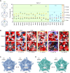
The Structural Diversity of Encapsulin Protein Shells
- PMID: 39330624
- PMCID: PMC11664910
- DOI: 10.1002/cbic.202400535
The Structural Diversity of Encapsulin Protein Shells
Abstract
Subcellular compartmentalization is a universal feature of all cells. Spatially distinct compartments, be they lipid- or protein-based, enable cells to optimize local reaction environments, store nutrients, and sequester toxic processes. Prokaryotes generally lack intracellular membrane systems and usually rely on protein-based compartments and organelles to regulate and optimize their metabolism. Encapsulins are one of the most diverse and widespread classes of prokaryotic protein compartments. They self-assemble into icosahedral protein shells and are able to specifically internalize dedicated cargo enzymes. This review discusses the structural diversity of encapsulin protein shells, focusing on shell assembly, symmetry, and dynamics. The properties and functions of pores found within encapsulin shells will also be discussed. In addition, fusion and insertion domains embedded within encapsulin shell protomers will be highlighted. Finally, future research directions for basic encapsulin biology, with a focus on the structural understand of encapsulins, are briefly outlined.
Keywords: Encapsulin; HK97; Nanocompartment; Protein capsid; Protein shell.
© 2024 The Author(s). ChemBioChem published by Wiley-VCH GmbH.
Conflict of interest statement
The authors declare no conflict of interest.
Figures


Similar articles
-
Encapsulins.Annu Rev Biochem. 2022 Jun 21;91:353-380. doi: 10.1146/annurev-biochem-040320-102858. Epub 2022 Mar 18. Annu Rev Biochem. 2022. PMID: 35303791 Free PMC article. Review.
-
Structure and heterogeneity of a highly cargo-loaded encapsulin shell.J Struct Biol. 2023 Dec;215(4):108022. doi: 10.1016/j.jsb.2023.108022. Epub 2023 Aug 30. J Struct Biol. 2023. PMID: 37657675 Free PMC article.
-
A Two-Component Pseudo-Icosahedral Protein Nanocompartment with Variable Shell Composition and Irregular Tiling.Adv Sci (Weinh). 2025 Aug;12(32):e03617. doi: 10.1002/advs.202503617. Epub 2025 Jun 25. Adv Sci (Weinh). 2025. PMID: 40557621 Free PMC article.
-
Encapsulins: molecular biology of the shell.Crit Rev Biochem Mol Biol. 2017 Oct;52(5):583-594. doi: 10.1080/10409238.2017.1337709. Epub 2017 Jun 21. Crit Rev Biochem Mol Biol. 2017. PMID: 28635326 Review.
-
Assembly and Mechanical Properties of the Cargo-Free and Cargo-Loaded Bacterial Nanocompartment Encapsulin.Biomacromolecules. 2016 Aug 8;17(8):2522-9. doi: 10.1021/acs.biomac.6b00469. Epub 2016 Jul 7. Biomacromolecules. 2016. PMID: 27355101
Cited by
-
In situ and in vitro cryo-EM reveal structures of mycobacterial encapsulin assembly intermediates.Commun Biol. 2025 Feb 15;8(1):245. doi: 10.1038/s42003-025-07660-5. Commun Biol. 2025. PMID: 39955411 Free PMC article.
-
Engineering encapsulin nanocages for drug delivery.Mater Adv. 2025 Jul 18. doi: 10.1039/d5ma00386e. Online ahead of print. Mater Adv. 2025. PMID: 40717769 Free PMC article. Review.
-
Controlling nanocage assembly, towards developing a one-health "plug & play" platform for targeted therapy.Chem Commun (Camb). 2025 Aug 28;61(71):13221-13235. doi: 10.1039/d5cc03592a. Chem Commun (Camb). 2025. PMID: 40824119 Free PMC article. Review.
-
Structural and Biochemical Characterization of a Widespread Enterobacterial Peroxidase Encapsulin.Adv Sci (Weinh). 2025 Jun;12(21):e2415827. doi: 10.1002/advs.202415827. Epub 2025 Apr 1. Adv Sci (Weinh). 2025. PMID: 40167211 Free PMC article.
References
-
- Diekmann Y., Pereira-Leal J. B., Biochem. J. 2013, 449, 319–331. - PubMed
-
- Aussignargues C., Pandelia M. E., Sutter M., Plegaria J. S., Zarzycki J., Turmo A., Huang J., Ducat D. C., Hegg E. L., Gibney B. R., Kerfeld C. A., J. Am. Chem. Soc. 2016, 138, 5262–5270. - PubMed
-
- Kerfeld C. A., Heinhorst S., Cannon G. C., Annu. Rev. Microbiol. 2010, 64, 391–408. - PubMed
Publication types
MeSH terms
Substances
Grants and funding
LinkOut - more resources
Full Text Sources

