Review
. 2024 Sep 1;10(9):1365-1378.
doi: 10.3390/tomography10090103.

Diagnostic and Therapeutic Approach to Thoracic Outlet Syndrome

Affiliations
Review

Diagnostic and Therapeutic Approach to Thoracic Outlet Syndrome

Stefania Rizzo et al. Tomography. .

Abstract

Thoracic outlet syndrome (TOS) is a group of symptoms caused by the compression of neurovascular structures of the superior thoracic outlet. The knowledge of its clinical presentation with specific symptoms, as well as proper imaging examinations, ranging from plain radiographs to ultrasound, computed tomography and magnetic resonance imaging, may help achieve a precise diagnosis. Once TOS is recognized, proper treatment may comprise a conservative or a surgical approach.

Keywords: CT; MRI; US; diagnosis; surgery; thoracic outlet syndrome; treatment.


Conflict of interest statement

The authors declare no conflict of interest.

Figures

Figure 1
Figure 1
MRI disclosing left subclavian vein stenosis (white arrow) after ipsilateral arm abduction.
Figure 2
Figure 2
CT scan disclosing left subclavian artery compression by complete supernumerary cervical rib (white arrow).
Figure 3
Figure 3
Intraoperative view of subclavian artery prosthetic reconstruction.
Figure 4
Figure 4
CT scan disclosing bilateral anterior scalene muscle hypertrophy (white arrows).
Figure 5
Figure 5
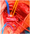
Intraoperative view of scalenotomy: upper blue vessel loop: superior trunk of the brachial plexus (C5 and C6 roots); yellow vessel loop: middle trunk of the brachial plexus (C7 root); red vessel loop: lower brachial plexus trunk (C8 and T1 roots).

References

    1. Panther E.J., Reintgen C.D., Cueto R.J., Hao K.A., Chim H., King J.J. Thoracic outlet syndrome: A review. J. Shoulder Elbow Surg. 2022;31:e545–e561. doi: 10.1016/j.jse.2022.06.026. - DOI - PubMed
    1. Illig K.A., Donahue D., Duncan A., Freischlag J., Gelabert H., Johansen K., Jordan S., Sanders R., Thompson R. Reporting standards of the Society for Vascular Surgery for thoracic outlet syndrome. J. Vasc. Surg. 2016;64:e23–e35. doi: 10.1016/j.jvs.2016.04.039. - DOI - PubMed
    1. Khalilzadeh O., Glover M., Torriani M., Gupta R. Imaging Assessment of Thoracic Outlet Syndrome. Thorac. Surg. Clin. 2021;31:19–25. doi: 10.1016/j.thorsurg.2020.09.002. - DOI - PubMed
    1. Wilbourn A.J. 10 most commonly asked questions about thoracic outlet syndrome. Neurologist. 2001;7:309–312. doi: 10.1097/00127893-200109000-00008. - DOI - PubMed
    1. Altuwaijri T.A. Comparison of duplex ultrasound and hemodynamic assessment with computed tomography angiography in patients with arterial thoracic outlet syndrome. Medicine. 2022;101:e30360. doi: 10.1097/MD.0000000000030360. - DOI - PMC - PubMed

MeSH terms

LinkOut - more resources