. 2024 Sep 3;11(9):407.
doi: 10.3390/vetsci11090407.

Does Catheter Insertion Site Matter? Contamination of Peripheral Intravenous Catheters during Dental Scaling in Dogs

Affiliations

Does Catheter Insertion Site Matter? Contamination of Peripheral Intravenous Catheters during Dental Scaling in Dogs

Ivana Calice et al. Vet Sci. .

Abstract

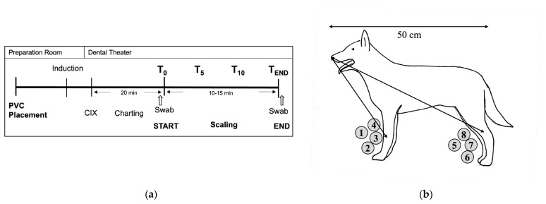
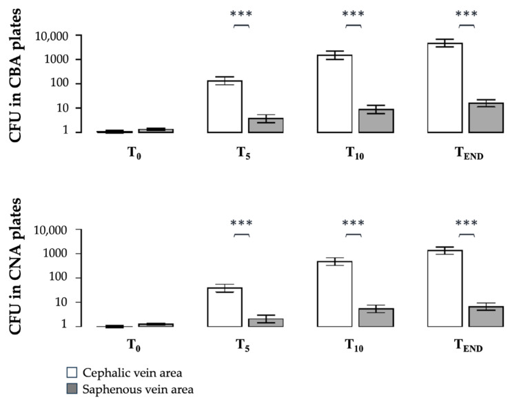
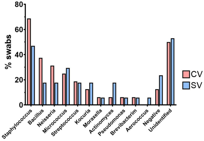
During dental scaling in dogs under general anaesthesia, contamination of the peripheral intravenous catheter (PIVC) is unavoidable due to splatter and the generated aerosol. Bacterial contamination was compared between two commonly used PIVC placement sites. Thirty-nine client-owned dogs with a minimum length from their nose to their tail base of 50 cm were randomly assigned to receive a PIVC in either their cephalic or saphenous vein. Irrespective of the PIVC placement site, brain heart infusion agar dishes were placed in the cephalic and saphenous vein areas. Their lids were closed 0, 5, and 10 min into the procedure. Contamination was measured by counting the colony-forming units after incubation on different substrates. The data were analysed with descriptive statistics, ANOVA, and ANCOVA (p < 0.05). The cephalic vein area showed a significantly higher bacterial load than the saphenous vein area (p ≈ 0.0) regardless of the length of the dog. Furthermore, the dorsal PIVC injection ports were sampled before and after scaling, and the colonies isolated were counted and subjected to MALDI-TOF-MS for identification. The bacteria mainly belonged to the genera Staphylococcus, Neisseria, and Bacillus. Our results suggest that for dental scaling in dogs, the PIVC should be placed in the pelvic limb whenever possible to reduce the potential risk of contamination.

Keywords: cannula; contamination; dental scaling; dog; peripheral intravenous catheter.


Conflict of interest statement

The authors declare no conflicts of interest.

Figures

Figure 1
Figure 1
(a) Study protocol timeline. CIX: chlorhexidine; Induction: induction of general anaesthesia; T0, T5, T10, TEND: time points for closure of sedimentation plates (study part A); Swab: time of dorsal peripheral intravenous catheter (PIVC) injection port swabbing (start and the end of the procedure, study part B). (b) Schematic drawing of the experimental setup of a dog positioned for dental scaling: the arrows show how the distance from the nose to the PIVC placement sites was measured for the cephalic vein (CV) and the saphenous vein (SV) and the positions of the sedimentation plates (grey and labelled 1–8); double-headed arrow above the dog shows how was the length of the dog measured (from nose to base of the tail).
Figure 2
Figure 2
Results of part A. Number of colony-forming units (CFU) per time point (T0, T5, T10, TEND) on CBA (clindamycin blood agar, selective for Gram-negative bacteria) and CNA (Columbia naladixicacid agar, selective for the growth of Gram-positive bacteria) at the cephalic vein (CV) (light columns) and the saphenous vein (SV) (dark columns), measured during dental scaling in dogs (n = 39). Data are presented as mean ± standard error of the log-transformed data. The average of the CFU counted per time point and allocation (CV vs. SV) was used for the statistical analysis (one-way ANOVA). Significance was set at p < 0.05. *** CFU differ significantly (p < 0.0001) between the CV and the SV at the measured time point.
Figure 3
Figure 3
Results part A. Scatterplot of distance from the nose to the cephalic (open circles) and saphenous vein (crosses) areas of the dogs (n = 39) during dental scaling on the x-axis and bacterial colony-forming unit (CFU) counts of the CBA (Gram-negative-selective agar) plates on the y-axis at time point 10 (10 min into dental scaling); grey lines connect the two samples belonging to the same dog. With an increasing distance from the mouth, the CFU counts decreased (T10), except in 4 animals.
Figure 4
Figure 4
Results of part B. MALDI-TOF-MS analysis of the most frequently isolated genera from the swabs sampled from the dorsal PIVC injection ports of the dogs during dental scaling. CV: cephalic vein (n = 18); SV: saphenous vein (n = 21); %swabs: percentage of swabs with isolated bacterial species.

Similar articles

References

    1. Timmerman M.F., Menso L., Steinfort J., Winkelhoff A.J.V., Weijden G.A.V.D. Atmospheric Contamination during Ultrasonic Scaling. J. Clin. Periodontol. 2004;31:458–462. doi: 10.1111/j.1600-051X.2004.00511.x. - DOI - PubMed
    1. Veena H.R., Mahantesha S., Joseph P.A., Patil S.R., Patil S.H. Dissemination of Aerosol and Splatter during Ultrasonic Scaling: A Pilot Study. J. Infect. Public Health. 2015;8:260–265. doi: 10.1016/j.jiph.2014.11.004. - DOI - PubMed
    1. Leggat P.A., Kedjarune U. Bacterial Aerosols in the Dental Clinic: A Review. Int. Dent. J. 2001;51:39–44. doi: 10.1002/j.1875-595X.2001.tb00816.x. - DOI - PubMed
    1. Jones I.D., Case A.M., Stevens K.B., Boag A., Rycroft A.N. Factors Contributing to the Contamination of Peripheral Intravenous Catheters in Dogs and Cats. Vet. Rec. 2009;164:616. doi: 10.1136/vr.164.20.616. - DOI - PubMed
    1. Mathews K.A., Brooks M.J., Valliant A.E. A Prospective Study Of Intravenous Catheter Contamination. J. Vet. Emerg. Crit. Care. 1996;6:33–43. doi: 10.1111/j.1476-4431.1996.tb00032.x. - DOI

LinkOut - more resources