. 2024 Oct;121(40):e2404243121.
doi: 10.1073/pnas.2404243121. Epub 2024 Sep 27.

Subcellular activation of β-adrenergic receptors using a spatially restricted antagonist

Affiliations

Subcellular activation of β-adrenergic receptors using a spatially restricted antagonist

Federica Liccardo et al. Proc Natl Acad Sci U S A. 2024 Oct.

Abstract

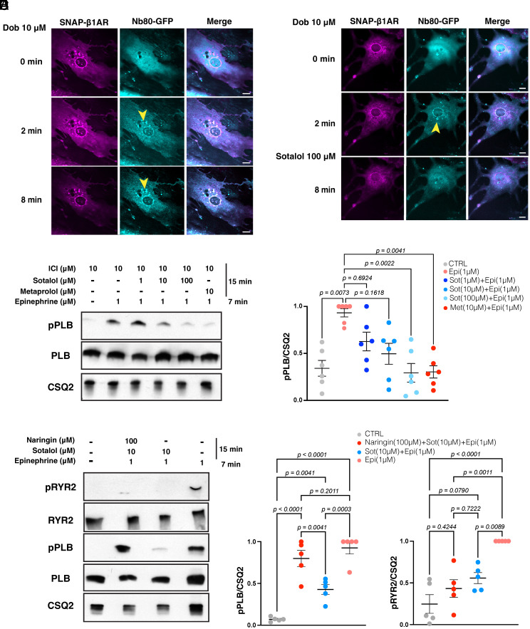
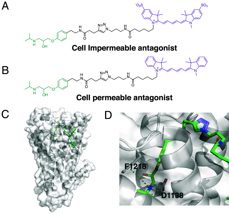
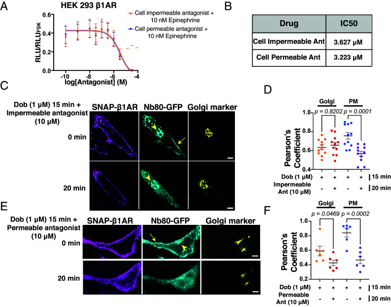
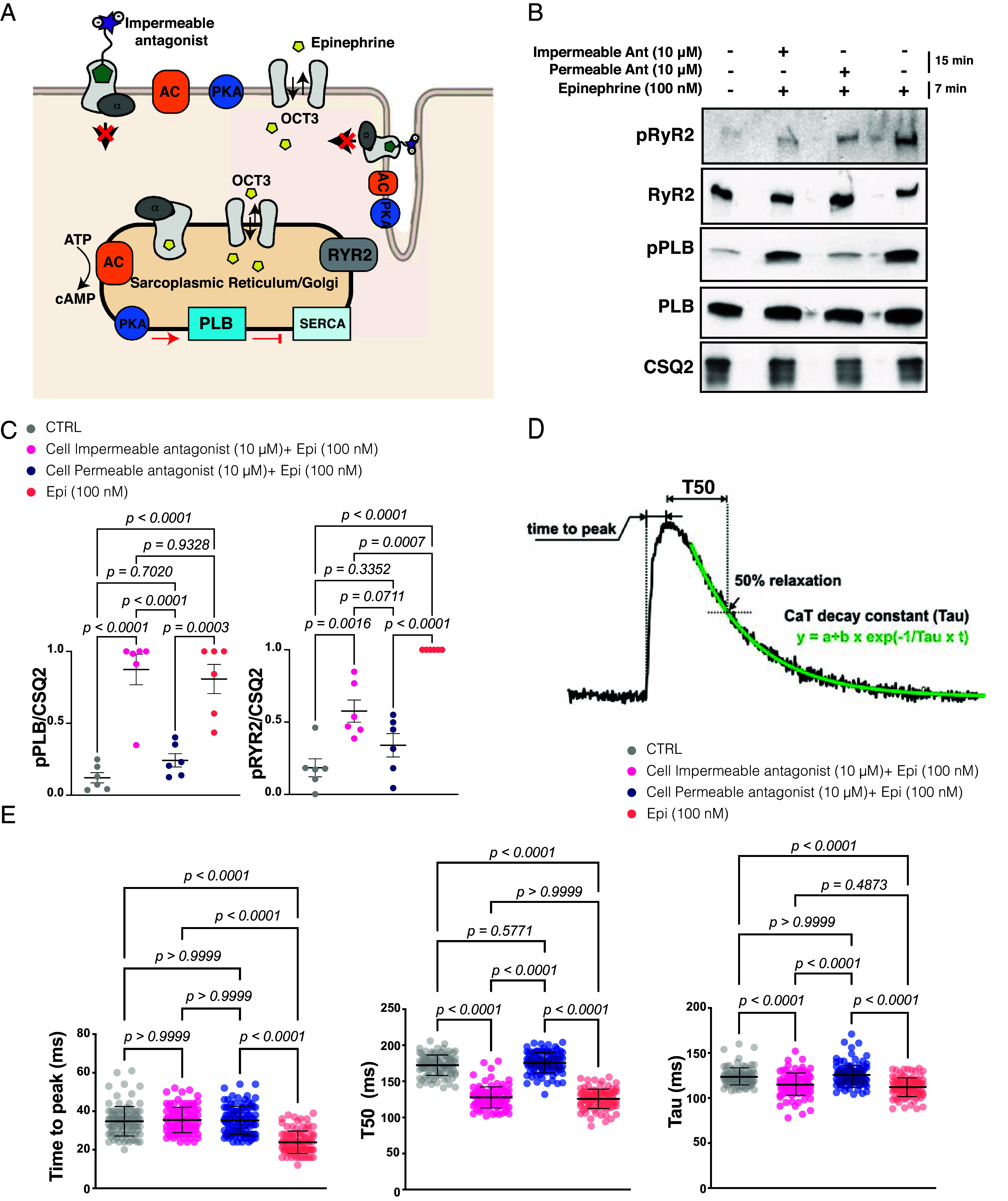
Gprotein-coupled receptors (GPCRs) regulate several physiological and pathological processes and represent the target of approximately 30% of Food and Drug Administration-approved drugs. GPCR-mediated signaling was thought to occur exclusively at the plasma membrane. However, recent studies have unveiled their presence and function at subcellular membrane compartments. There is a growing interest in studying compartmentalized signaling of GPCRs. This requires development of tools to separate GPCR signaling at the plasma membrane from the ones initiated at intracellular compartments. We leveraged the structural and pharmacological information available for β-adrenergic receptors (βARs) and focused on β1AR as exemplary GPCR that functions at subcellular compartments, and rationally designed spatially restricted antagonists. We generated a cell-impermeable βAR antagonist by conjugating a suitable pharmacophore to a sulfonate-containing fluorophore. This cell-impermeable antagonist only inhibited β1AR on the plasma membrane. In contrast, a cell-permeable βAR antagonist containing a nonsulfonated fluorophore efficiently inhibited both the plasma membrane and Golgi pools of β1ARs. Furthermore, the cell-impermeable antagonist selectively inhibited the phosphorylation of PKA downstream effectors near the plasma membrane, which regulate sarcoplasmic reticulum (SR) Ca2+ release in adult cardiomyocytes, while the β1AR Golgi pool remained active. Our tools offer promising avenues for investigating compartmentalized βAR signaling in various contexts, potentially advancing our understanding of βAR-mediated cellular responses in health and disease. They also offer a general strategy to study compartmentalized signaling for other GPCRs in various biological systems.

Keywords: GPCR signaling; Pharmacology; drug design.


Conflict of interest statement

Competing interests statement:K.M.S. has consulting agreements for the following companies, which involve monetary and/or stock compensation: Revolution Medicines, Black Diamond Therapeutics, BridGene Biosciences, Denali Therapeutics, Dice Molecules, eFFECTOR Therapeutics, Erasca, Genentech/Roche, Janssen Pharmaceuticals, Kumquat Biosciences, Kura Oncology, Mitokinin, Nested, Type6 Therapeutics, Venthera, Wellspring Biosciences (Araxes Pharma), Turning Point, Ikena, Initial Therapeutics, Vevo and BioTheryX.

Figures

Fig. 1.
Fig. 1.
Sotalol inhibits both the plasma membrane and the Golgi-localized β1AR in cardiomyocytes. (A) Representative confocal images of NCMs expressing the conformational biosensor of β1AR, Nb80-GFP (cyan), and SNAP-β1AR (magenta) before and after dobutamine (10 μM) treatment for 10 min. Stimulation with dobutamine (10 μM) results in Nb80-GFP recruitment to the plasma membrane and the Golgi. Arrowheads indicate Nb80-GFP localization at the Golgi. (Scale bar: 10 μm.) (B) Representative confocal images of NCMs expressing Nb80-GFP (cyan) and SNAP-β1AR (magenta) pretreated with dobutamine (10 μM) and before and after sotalol (100 μM) addition. Sotalol (100 μM) treatment for 3 min results in the loss of Nb80-GFP localization at the Golgi. Arrowheads indicate Nb80-GFP localization at the Golgi. (Scale bar: 10 μm.) (C) Representative phosphorylation profile of PLB (pPLB) regulated by β1AR in adult cardiomyocytes (ACM) derived from wild-type mice. ACM were pretreated with 10 μM β2AR-selective antagonist ICI-118551 (ICI) to isolate the function of β1AR. Phosphorylation of PLB Ser16/Thr17 (pPLB) was analyzed in wild-type ACM upon treatment with metoprolol (10 μM) or sotalol (1, 10, and 100 μM) for 15 min and followed by epinephrine (1 μM) treatment for 7 min at 37 °C. (D) Quantification of immunoblots of pPLB was normalized to the protein levels of CSQ2 and then reported as a percentage of the highest value in the groups. The quantified data from different experiments are presented as mean ± SEM. The P-values were calculated by two-way ANOVA. n = 6 independent biological replicates. (E) Representative phosphorylation profile of RyR2 (pRyR2) and PLB (pPLB) in ACM derived from wild-type mice. Phosphorylation of RyR2 Ser2808 (pRyR2) and PLB Ser16/Thr17 (pPLB) were analyzed upon treatment with OATP1A2-selective inhibitor Naringin (100 μM) for 15 min, sotalol (10 μM) for 15 min followed by epinephrine (1 μM) treatment for 7 min at 37 °C. (F) Quantification of immunoblots of pRyR2 and pPLB was normalized to the protein levels of CSQ2 and then reported as a percentage of the highest value in the groups. The quantified data from different experiments are presented as mean ± SEM. The P-values were calculated by two-way ANOVA. n = 5 independent biological replicates.
Fig. 2.
Fig. 2.
Rational design and synthesis of spatially restricted βAR antagonists. (A) Chemical structures of cell-impermeable antagonist. Pharmacophore moiety of the antagonist (green) is functionalized with a linker (black) and a fluorophore (purple). The cell-impermeable antagonist is conjugated with a sulfonated Cy5. (B) Chemical structures of cell-permeable antagonist. Pharmacophore moiety of the antagonist (green) is functionalized with a linker (black) and a fluorophore (purple). The cell-permeable antagonist is conjugated with a Cy5 dye. (C) Computational analysis of the binding interaction between cell-permeable antagonist and β1AR (PDB:7BTS). (D) β1AR orthosteric pocket and pharmacophore interaction sites. D1138 and F1218 are β1AR residues interacting with the pharmacophore via H-bond an π-cation interaction, respectively.
Fig. 3.
Fig. 3.
Conformational biosensor, Nb80-GFP, detects activated β1AR before and after cell-permeable or -impermeable antagonists’ treatment. (A) Concentration–response curve of the cell-permeable (blue line) and the cell-impermeable (orange line) antagonists in HEK293 overexpressing β1AR and a bioluminescent cAMP biosensor. Cells were treated with different concentrations of antagonists in combination with ∼10 nM epinephrine. Relative luminescence values are normalized to HEK293 cells treated with forskolin (20 μM). n = 4 independent biological replicates. (B) IC50 values of cell-impermeable and -permeable antagonist are respectively 3.627 μM and 3.223 μM. (C) Confocal images of representative HeLa cells expressing Nb80-GFP (cyan) and SNAP-β1AR (SNAP surface staining, magenta) and the Golgi marker (yellow), pretreated with dobutamine (1 μM) for 15 min. Stimulation with cell-impermeable antagonist (10 μM) for 20 min selectively inhibits the plasma membrane-localized β1AR. The arrow indicates the plasma membrane, and arrowheads indicate Golgi localization. (Scale bar: 10 μm.) (D) Person’s correlation coefficient measurements for the colocalization analysis of Nb80-GFP at the Golgi and plasma membrane after treatments of cardiomyocytes with the cell-impermeable antagonist. The results have been measured from n = 10 cells, three independent biological replicates; P values were calculated by one-way ANOVA. (E) Confocal images of representative HeLa cells expressing Nb80-GFP (cyan) and SNAP-β1AR (SNAP surface staining, magenta) and the Golgi marker (yellow), pretreated with dobutamine (1 μM) for 15 min. Stimulation with cell-permeable antagonist (10 μM) for 20 min inhibits both the plasma membrane and the Golgi pool of β1AR. The arrow indicates the plasma membrane, and arrowheads indicate Golgi localization. (Scale bar: 10 μm.) (F) Person’s correlation coefficient measurements for the colocalization analysis of Nb80-GFP at the Golgi and plasma membrane for cardiomyocytes treated with the cell-permeable antagonist. The results have been measured from n = 6 cells, two independent biological replicates; P-values were calculated by one-way ANOVA.
Fig. 4.
Fig. 4.
Cell-impermeable antagonist only inhibits β1AR-mediated responses from the plasma membrane. (A) Illustration of the effect of membrane-impermeable antagonist in blocking β1AR-mediated signaling in ACM. (B) Representative phosphorylation profiles of RyR2 and PLB induced by epinephrine (100 nM) alone, or in combination with the cell-impermeable and -permeable antagonists (10 μM) in ACM. Phosphorylation of RyR2 Ser2808 (pRyR2) and PLB Ser16/Thr17 (pPLB) was analyzed in wild-type ACMs endogenously expressing β1AR by immunoblotting. (C) Quantification of immunoblots of pPLB and pRYR2 was normalized to the protein levels of CSQ2 and then reported as a percentage of the highest value in the groups. The quantified data from different experiments are presented as mean ± SEM. The P-values were calculated by two-way ANOVA. n = 6 independent biological replicates. (D) Representative Ca2+ transient trace analysis. (E) Comprehensive quantification and comparison of time to peak (TTP), T50, and decay constant (Tau) from the four groups. N = 100 cells per condition in n = 3 independent biological replicates. One-way ANOVA was used to compare the parameters with the post hoc test using Bonferroni’s multiple comparison test. The data are presented as mean± SD.

Update of

Similar articles

Cited by

References

    1. Pierce K. L., Premont R. T., Lefkowitz R. J., Seven-transmembrane receptors. Nat. Rev. Mol. Cell Biol. 3, 639–650 (2002). - PubMed
    1. Calebiro D., et al. , Persistent cAMP-signals triggered by internalized G-protein-coupled receptors. PLoS Biol. 7, e1000172 (2009). - PMC - PubMed
    1. Feinstein T. N., et al. , Noncanonical control of vasopressin receptor type 2 signaling by retromer and arrestin. J. Biol. Chem. 288, 27849–27860 (2013). - PMC - PubMed
    1. Ferrandon S., et al. , Sustained cyclic AMP production by parathyroid hormone receptor endocytosis. Nat. Chem. Biol. 5, 734–742 (2009). - PMC - PubMed
    1. Kotowski S. J., Hopf F. W., Seif T., Bonci A., von Zastrow M., Endocytosis promotes rapid dopaminergic signaling. Neuron 71, 278–290 (2011). - PMC - PubMed