. 2024 Oct;121(40):e2318098121.
doi: 10.1073/pnas.2318098121. Epub 2024 Sep 27.

Poly ADP-ribose signaling is dysregulated in Huntington disease

Affiliations

Poly ADP-ribose signaling is dysregulated in Huntington disease

Tamara Maiuri et al. Proc Natl Acad Sci U S A. 2024 Oct.

Abstract

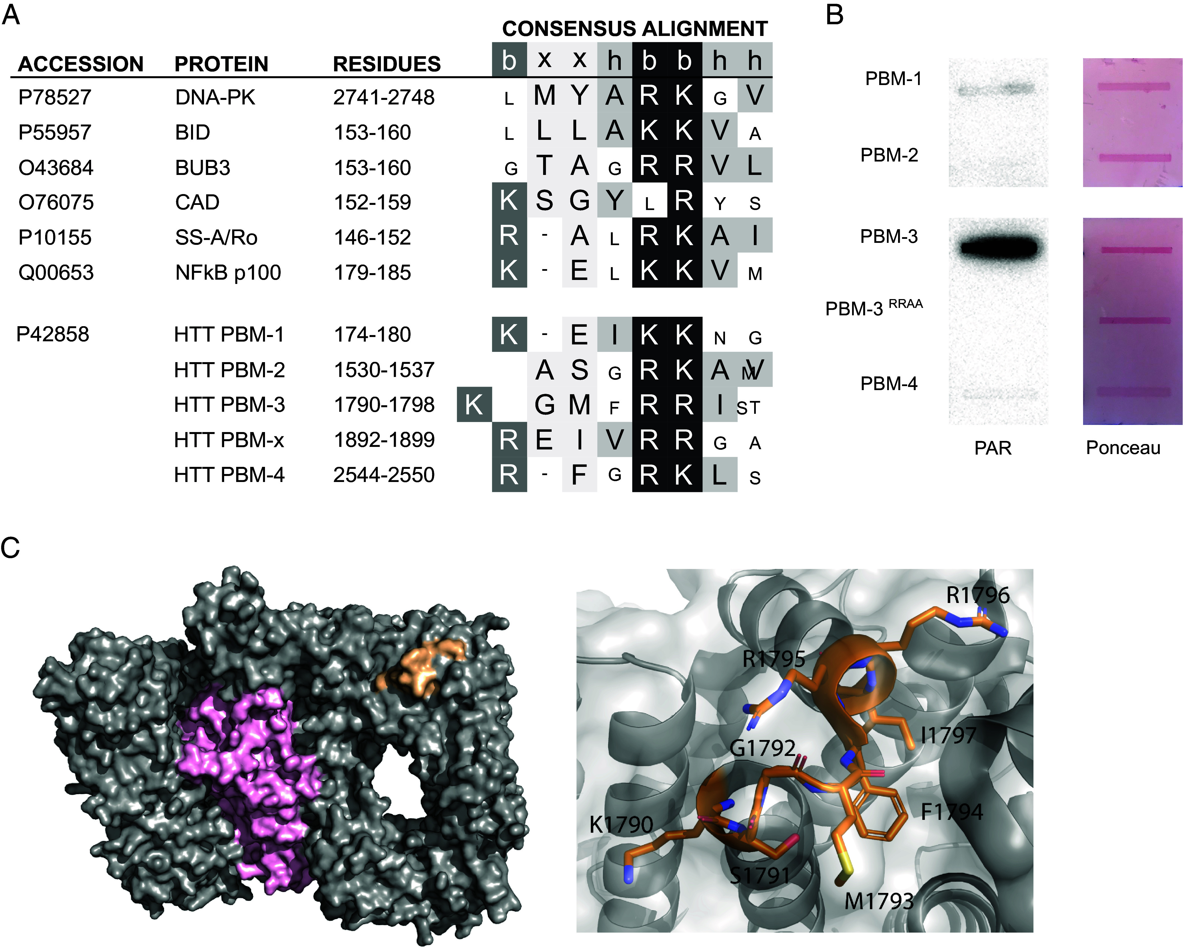
Huntington disease (HD) is a genetic neurodegenerative disease caused by cytosine, adenine, guanine (CAG) expansion in the Huntingtin (HTT) gene, translating to an expanded polyglutamine tract in the HTT protein. Age at disease onset correlates to CAG repeat length but varies by decades between individuals with identical repeat lengths. Genome-wide association studies link HD modification to DNA repair and mitochondrial health pathways. Clinical studies show elevated DNA damage in HD, even at the premanifest stage. A major DNA repair node influencing neurodegenerative disease is the PARP pathway. Accumulation of poly adenosine diphosphate (ADP)-ribose (PAR) has been implicated in Alzheimer and Parkinson diseases, as well as cerebellar ataxia. We report that HD mutation carriers have lower cerebrospinal fluid PAR levels than healthy controls, starting at the premanifest stage. Human HD induced pluripotent stem cell-derived neurons and patient-derived fibroblasts have diminished PAR response in the context of elevated DNA damage. We have defined a PAR-binding motif in HTT, detected HTT complexed with PARylated proteins in human cells during stress, and localized HTT to mitotic chromosomes upon inhibition of PAR degradation. Direct HTT PAR binding was measured by fluorescence polarization and visualized by atomic force microscopy at the single molecule level. While wild-type and mutant HTT did not differ in their PAR binding ability, purified wild-type HTT protein increased in vitro PARP1 activity while mutant HTT did not. These results provide insight into an early molecular mechanism of HD, suggesting possible targets for the design of early preventive therapies.

Keywords: Huntington’s disease; PARP1; PARylation; huntingtin; poly ADP-ribose.


Conflict of interest statement

Competing interests statement:E.J.W. reports consultancy/ advisory board memberships with Annexon, Remix Therapeutics, Hoffman La Roche Ltd., Ionis Pharmaceuticals, PTC Therapeutics, Takeda, Teitur Trophics, Triplet Therapeutics and Vico Therapeutics. All honoraria for these consultancies were paid through the offices of UCL Consultants Ltd., a wholly owned subsidiary of University College London. F.B.R. is a Medpace UK Ltd. employee and was a University College London employee during the conduct of this study. F.B.R. has provided consultancy services to GLG and F. Hoffmann-La Roche Ltd. L.M.B. currently holds consultancy contracts with Annexon Biosciences, Remix Therapeutics, and LoQus23 Therapeutics Ltd. via UCL Consultants Ltd. R.T. has past consultancy with Novartis AG, PTC Therapeutics and Mitokinin LLC.

Figures

Fig. 1.
Fig. 1.
PAR levels are reduced in premanifest and manifest HD patient CSF. CSF samples from control, premanifest HD, and manifest HD subjects were blinded and analyzed for PAR levels by ELISA. Group comparisons were assessed using multiple regression with post hoc Wald tests. *Survives Bonferroni correction for multiple comparisons.
Fig. 2.
Fig. 2.
The PAR response is deficient in HD cells. (A) iPSC-derived neurons were fixed and stained with neuronal marker Map2 (red) and MABE1031 PAR detection reagent (green). Nuclear PAR intensity in Map2-positive cells was measured using CellProfiler. Data from six (CTR Q18 and HD Q53) or four (JHD Q77) differentiation replicates are shown (n = 100 to 300 nuclei per cell line). Results were analyzed by the Kruskal–Wallis test and corrected for multiple comparisons using Dunn's test. Error bars: SEM. (B) hTERT-immortalized fibroblasts from healthy control (TruHD-Q21Q18) and HD patients (TruHD-Q43Q17 and TruHD-Q50Q40) were treated with 100 mM KBrO3 for 30 min followed by Repair Assisted Damage Detection (RADD) to detect DNA damage, and costaining with MABE1031 PAR detection reagent. Representative images of TruHD-Q21Q18 cells are shown. Nuclear RADD and PAR intensity were measured using CellProfiler, mean intensity recorded for each image (18 images per condition; >500 cells), and values normalized to the control condition. Data from three independent experiments are shown. Results were analyzed by two-way ANOVA and corrected for multiple comparisons using Tukey’s test. Error bars: SEM. (Scale bars: 50 microns.)
Fig. 3.
Fig. 3.
PARP1/2 activity is higher in the presence of wild-type HTT. Fibroblasts were pretreated with increasing doses of PDD00017273 (A) or veliparib (B) for 30 min followed by 100 mM KBrO3 for 30 min in the presence of inhibitor. Veliparib dose–response was carried out in the presence of 5 μM PARG inhibitor to enable pan-ADP-ribose detection by MABE1016. EC50 and IC50 values were calculated from nuclear PAR staining intensity (10 to 12 images per condition; >800 cells) using GraphPad Prism. Error bars = SEM for four (PARG inhibitor) or eight (veliparib) experiments. ****P < 0.0001 (Brown–Forsythe and Welch ANOVA tests). (C) 10 fmol recombinant PARP1 was incubated with the indicated amounts of recombinant HTT-HAP40 for 2 h at 30 °C. Reactions were separated by SDS-PAGE and immunoblotted with MABE1016 pan-ADP-ribose detection reagent. Signal intensities were quantified using ImageJ. Values from three (HTT-HAP40 Q54) or four (HTT-HAP40 Q23) experiments are shown.
Fig. 4.
Fig. 4.
HTT interacts with PARylated proteins. (A) Degree of overlap between HTT interacting proteins and a list of PARylated proteins compiled from three independent studies, with Fisher’s exact test for statistical significance. (B) RPE1 cells were treated with 400 μM H2O2 in HBSS for 10 min and proteins cross-linked with 1% paraformaldehyde prior to lysis. HTT was immunoprecipitated with EPR5526 and associated proteins separated by SDS-PAGE and immunoblotted with the indicated antibodies. PARylated proteins of various sizes in the whole cell lysate (input) and anti-HTT IP were detected with pan ADP-ribose detection reagent (MABE1016) followed by HRP-conjugated anti-rabbit secondary antibody. Rabbit IgG signal from the anti-HTT immunoprecipitating antibody (EPR5526) was visible upon incubation with secondary anti-rabbit antibody. Results representative of four experiments. (C) RPE1 cells were treated with either 10 μM PDD00017273 PARG inhibitor (Top) or 1 μM talazoparib PARP1/2 inhibitor (Bottom) for 40 min prior to methanol fixation and immunofluorescence against HTT phosphorylated at residues S13 and S16 within the N17 domain (HTT phospho-N17, yellow), and PARP1 (PARP1, magenta), followed by counterstaining with Hoechst (DNA, cyan). Image representative of all mitotic cells observed (n > 10 cells from two independent experiments). (Scale bar: 10 μm.)
Fig. 5.
Fig. 5.
HTT has a PBM. (A) Known PBMs compared to putative PBMs in HTT. PBM-X is not solvent-accessible and was not analyzed further. b: basic, h: hydrophobic, x: any amino acid. Critical basic amino acids depicted in black boxes. (B) Peptides were slot-blotted onto nitrocellulose and then overlaid with 0.2 μM PAR polymer. After washing, anti-PAR western was performed with pan ADP-ribose detection reagent MABE1016. (C, Left) High-resolution cryoEM model of HTT–HAP40 complex (PDB–6X9O) shown in surface representation with HTT in gray, HAP40 in pink, and the PBM in orange. (C, Right) PBM shown in stick representation in orange. Positively charged K1790, R1795, and R1796 residues are surface exposed.
Fig. 6.
Fig. 6.
HTT directly binds PAR. (A) Fluorescence polarization assays using FAM-labeled 11-mer or 26-mer PAR and purified HTT-HAP40 Q23. Results for two experiments are shown. Error bars = SD. (B) Fluorescence polarization assays using FAM-labeled 26-mer PAR and 10 μM of the indicated subdomains of HTT. Reactions were carried out with 3 to 4 intra-assay replicates. Results for three experiments were analyzed by one-way ANOVA and corrected for multiple comparisons by the Kruskal–Wallis test (***P < 0.0005, ****P < 0.0001). (C) Recombinant HTT-HAP40 was added to PARP1 activity assays and reactions were deposited on mica and visualized by atomic force microscopy. 2D (Left) and 3D (Right) images are shown with corresponding color scale for PARylated PARP1 (2.0 nm) or PARylated PARP1 with HTT-HAP40 (10.0 nm).

References

    1. Lee J.-M., et al. , CAG repeat expansion in Huntington disease determines age at onset in a fully dominant fashion. Neurology 78, 690–695 (2012). - PMC - PubMed
    1. Genetic Modifiers of Huntington’s Disease (GeM-HD) Consortium, CAG repeat not polyglutamine length determines timing of Huntington’s disease onset. Cell 178, 887–900.e14 (2019). - PMC - PubMed
    1. Genetic Modifiers of Huntington’s Disease (GeM-HD) Consortium, Identification of genetic factors that modify clinical onset of Huntington’s disease. Cell 162, 516–526 (2015). - PMC - PubMed
    1. Djousse L., et al. , Interaction of normal and expanded CAG repeat sizes influences age at onset of Huntington disease. Am. J. Med. Genet. A 119, 279–282 (2003). - PubMed
    1. Wexler N. S., et al. , Venezuelan kindreds reveal that genetic and environmental factors modulate Huntington’s disease age of onset. Proc. Natl. Acad. Sci. U.S.A. 101, 3498–3503 (2004). - PMC - PubMed

MeSH terms

Substances