Review
. 2024 Oct 1;72(10):1424-1432.
doi: 10.4103/IJO.IJO_1549_23. Epub 2024 Sep 27.

Enhancing aesthetic outcomes: The role of biomechanics in periorbital and eyelid cosmetic surgery

Affiliations
Review

Enhancing aesthetic outcomes: The role of biomechanics in periorbital and eyelid cosmetic surgery

Lei Guo et al. Indian J Ophthalmol. .

Abstract

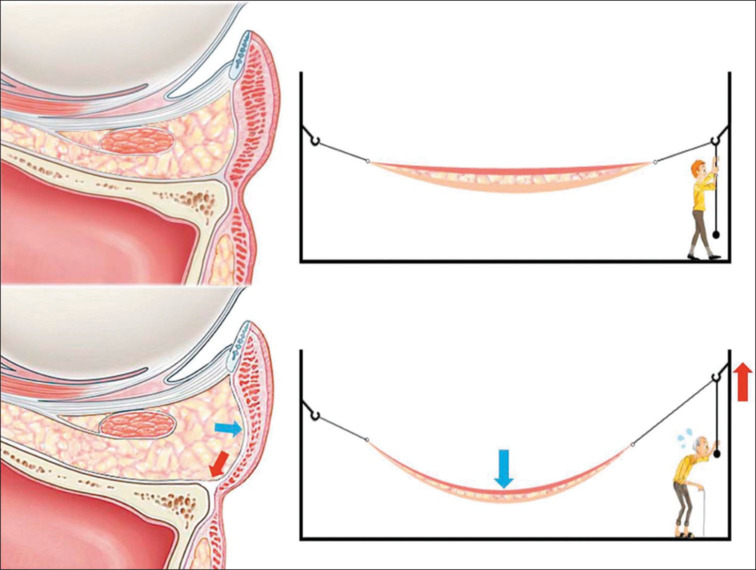
Cosmetic periorbital and eyelid surgery is a commonly performed procedure in facial plastic surgery. Understanding the biomechanics of periorbital anatomy and its role in aesthetic surgery is essential for achieving optimal outcomes. This review explores the biomechanical processes involved in periorbital age changes and analyzes the impact of cosmetic surgery approaches on these processes. By maintaining the initial mechanical equilibrium of the brows, eyelids, septal fat, and blepharoplasty folds, periorbital and eyelid cosmetic surgery can effectively rejuvenate the appearance. Disruption of this equilibrium can lead to the migration of anatomic components, resulting in signs of aging. Surgeons, by applying biomechanical concepts, can tailor the forces exerted upon the different structures to manifest the patient's aesthetic aspirations. The key to success in periorbital and eyelid cosmetic surgery lies in re-establishing a dynamic mechanical equilibrium within the periorbital framework.


Conflict of interest statement

There are no conflicts of interest.

Figures

Figure 1
Figure 1
Flow chart of conducted literature search
Figure 2
Figure 2
(Above) Youthful face. The muscle and gravitational forces that act on the anatomic structures of a young face are illustrated. (Below) Aged face. Characteristic changes that these forces produce in the older face (Reprinted with permission from Knize DM. Anatomic concepts for brow lift procedures. Plast Reconstr Surg. 2009;124(6):2118-2126.). STL = superior temporal fusion line of the skull
Figure 3
Figure 3
Attenuation of the levator aponeurosis with a high upper lid crease and ptotic lid margin (Reprinted with permission from Jindal K, Sarcia M, Codner MA. Functional considerations in aesthetic eyelid surgery. Plast Reconstr Surg. 2014;134(6):1154-1170.)
Figure 4
Figure 4
Diagram shows one of the methods of double blepharoplasty. (a) The sagittal plane demonstrates the modified Park’s method for double blepharoplasty, securing the orbicularis oculi muscle to the tarsal plate; (b) The coronal plane illustrates this method of double blepharoplasty, anchoring the orbicularis oculi muscle to the tarsal plate. (Reproduced with permission from Sun W, Wang Y, Song T, Wu D, Li H, Yin N. Orbicularis-tarsus fixation approach in double-eyelid blepharoplasty: A modification of Park’s technique. Aesthetic Plastic Surgery. 2018;42:1582-90.)
Figure 5
Figure 5
Diagram illustrating the mechanism for infraorbital fat herniation. The red arrow represents inferior displacement of the inferior orbital rim. The blue arrow represents dermal/fat/muscle atrophy. The aging man represents the loss of orbicularis oculi muscle tone. Mechanical stretching of the lower eyelid due to inferior displacement of the inferior orbital rim and weakening of the lower eyelid due to age-related factors such as dermal/fat/muscle atrophy and loss of muscle tone together make the lower eyelid droop anteriorly. Therefore, bulging herniation of the infraorbital fat can be thought of as a “hammock effect”(Reproduced with permission from Kim J, Park SW, Choi J, et al. Ageing of the bony orbit is a major cause of age-related intraorbital fat herniation. J Plast Reconstr Aesthet Surg. 2018;71(5):658-664.)
Figure 6
Figure 6
An illustration of a sagittal view of the lower lid showing the two main approaches to lower blepharoplasty. (a)The short dashed line represents the dissection plane of the lower blepharoplasty through the skin incision, while a dotted line represents the dissection plane of the lower blepharoplasty through the conjunctival incision. (b) In the transcutaneous lower blepharoplasty, the inferior oblique muscle is released, the retaining ligament is preserved, and the orbital fat is repositioned. (c) In transconjunctival lower eyelid blepharoplasty, the arcus marginalis is released simultaneously with the repositioning of the orbital fat.(Reproduced with permission from Alghoul M. Blepharoplasty: Anatomy, planning, techniques, and safety. Aesthetic Surgery Journal. 2019;39:10 28.)

Similar articles

References

    1. Ksander GA, Vistnes LM, Rose EH. Excisional wound biomechanics, skin tension lines, and elastic contraction. Plast Reconstr Surg. 1977;59:398–406. - PubMed
    1. Corr DT, Hart DA. Biomechanics of scar tissue and uninjured skin. Adv Wound Care (New Rochelle) 2013;2:37–43. - PMC - PubMed
    1. Fernandes MG, da Silva LP, Cerqueira MT, Ibañez R, Murphy CM, Reis RL, et al. Mechanomodulatory biomaterials prospects in scar prevention and treatment. Acta Biomater. 2022;150:22–33. - PubMed
    1. Hosseini M, Brown J, Khosrotehrani K, Bayat A, Shafiee A. Skin biomechanics: A potential therapeutic intervention target to reduce scarring. Burns Trauma. 2022;10:tkac036. - PMC - PubMed
    1. Furnas DW, Fischer GW. The Z-plasty: Biomechanics and mathematics. Br J Plast Surg. 1971;24:144–60. - PubMed

LinkOut - more resources