

Gasterophilus intestinalis infestation in lion (Panthera leo) and plains zebra (Equus quagga) in the Serengeti ecosystem: Morphological and molecular profiling
- PMID: 39331805
- PMCID: PMC11433831
- DOI: 10.1051/parasite/2024060
Gasterophilus intestinalis infestation in lion (Panthera leo) and plains zebra (Equus quagga) in the Serengeti ecosystem: Morphological and molecular profiling
Abstract
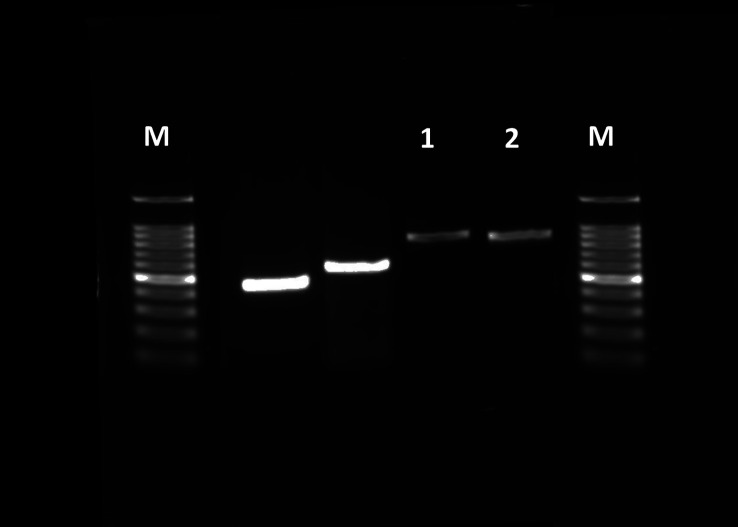
This study was conducted to clarify the host specificity and the geographical distribution of Gasterophilus species (Diptera, Oestridae) in the Serengeti ecosystem. A total of 317 larvae were recovered from two common zebras (Equus quagga, formerly Equus burchellii) in Maswa Game Reserve, and 58 larvae were recovered from an African lion (Panthera leo) in the Serengeti National Park. The study emphasizes the rare occurrence of Gasterophilus sp. in lions, shedding light on the broader life cycle and physiological implications for hosts. Genetic analysis of cox2 genes from Gasterophilus species, sourced from a single geographic location, reveals significant genetic distinctions and host specificity. This study reports the first case of G. intestinalis infestation in an African lion in the Serengeti ecosystem, extending its known range from zebras and other equids, and highlighting ecological and veterinary implications. This unusual prey-predator transmission highlights the value of molecular taxonomic tools in clarifying host-parasite dynamics and guiding targeted conservation strategies.
Title: Infestation par Gasterophilus intestinalis chez le lion (Panthera leo) et le zèbre des plaines (Equus quagga) dans l’écosystème du Serengeti : profilage morphologique et moléculaire.
Abstract: Cette étude a été menée pour clarifier la spécificité de l’hôte et la répartition géographique des espèces de Gasterophilus (Diptera, Oestridae) dans l’écosystème du Serengeti. Au total, 317 larves ont été récoltées chez deux zèbres communs (Equus quagga, anciennement Equus burchellii) dans la réserve de gibier de Maswa, et 58 larves ont été récoltées chez un lion d’Afrique (Panthera leo) dans le parc national du Serengeti. L’étude souligne la rareté de l’occurrence de Gasterophilus sp. chez les lions, mettant en lumière le cycle biologique plus large et les implications physiologiques pour les hôtes. L’analyse génétique des gènes cox2 des espèces de Gasterophilus, provenant d’un seul lieu géographique, révèle des distinctions génétiques et une spécificité d’hôte significatives. Cette étude rapporte le premier cas d’infestation par G. intestinalis chez un lion africain dans l’écosystème du Serengeti, étendant son aire de répartition déjà connue chez les zèbres et autres équidés, et mettant en évidence des implications écologiques et vétérinaires. Cette transmission inhabituelle de proie à prédateur souligne l’intérêt des outils de taxonomie moléculaire pour clarifier la dynamique hôte-parasite et guider les stratégies de conservation ciblées.
Keywords: Gasterophilus intestinalis; Lion; Maswa; Serengeti ecosystem; Zebra Tanzania.
© B. Abdieli Ndossi et al., published by EDP Sciences, 2024.
Conflict of interest statement
We have no conflicts of interests related to this work.
Figures


References
-
- Abdel-Rahman MM, Hassanen EA, Abdel Mageed MA. 2018. Light and scanning electron microscopy of Gasterophilus intestinalis (larvae and adult fly) infesting donkeys with emphasis on histopathology of the induced lesions. Egyptian Veterinary Medical Society of Parasitology Journal, 14(1), 15–31.
-
- Agneessens J, Engelen S, Debever P, Vercruysse J. 1998. Gasterophilus intestinalis infections in horses in Belgium. Veterinary Parasitology, 77(2–3), 199–204. - PubMed
-
- Akele Y, Enbiyale G, Negash A, Ayana E. 2018. Equine myiasis caused by Gastrophilus flies: a review. Acta Parasitologica Globalis, 9, 44–52.
-
- Attia MM, Salaeh NM. 2020. Ultrastructure of adult Gasterophilus intestinalis (Diptera: Gasterophilidae) and its puparium. International Journal of Tropical Insect Science, 40, 327–335.
-
- Bezdekova B, Jahn P, Vyskocil M. 2007. Pathomorphological study on gastroduodenal ulceration in horses: localisation of lesions. Acta Veterinaria Hungarica, 55(2), 241–249. - PubMed
MeSH terms
Substances
Grants and funding
LinkOut - more resources
Full Text Sources
Research Materials