Review
. 2025 Jan 1;31(1):21-47.
doi: 10.1093/humupd/dmae027.

New insights into the ovulatory process in the human ovary

Affiliations
Review

New insights into the ovulatory process in the human ovary

Misung Jo et al. Hum Reprod Update. .

Abstract

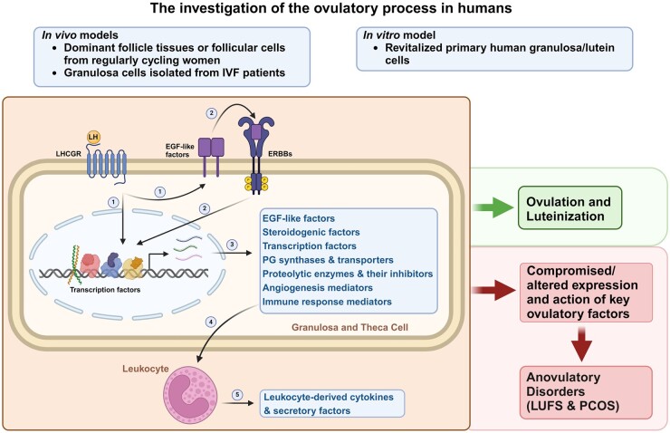
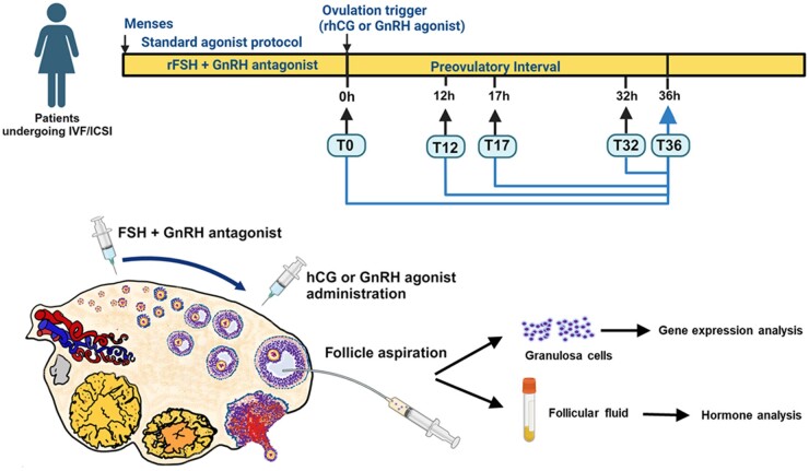
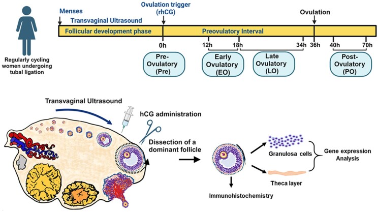
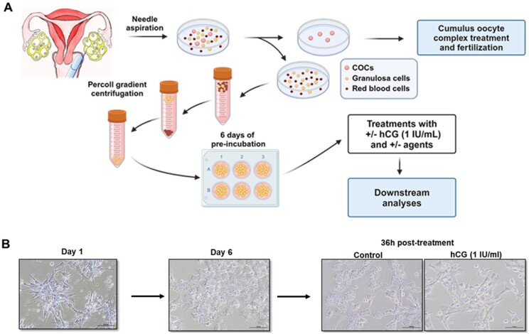
Background: Successful ovulation is essential for natural conception and fertility. Defects in the ovulatory process are associated with various conditions of infertility or subfertility in women. However, our understanding of the intra-ovarian biochemical mechanisms underlying this process in women has lagged compared to our understanding of animal models. This has been largely due to the limited availability of human ovarian samples that can be used to examine changes across the ovulatory period and delineate the underlying cellular/molecular mechanisms in women. Despite this challenge, steady progress has been made to improve our knowledge of the ovulatory process in women by: (i) collecting granulosa cells across the IVF interval, (ii) creating a novel approach to collecting follicular cells and tissues across the periovulatory period from normally cycling women, and (iii) developing unique in vitro models to examine the LH surge or hCG administration-induced ovulatory changes in gene expression, the regulatory mechanisms underlying the ovulatory changes, and the specific functions of the ovulatory factors.

Objective and rationale: The objective of this review is to summarize findings generated using in vivo and in vitro models of human ovulation, with the goal of providing new insights into the mechanisms underlying the ovulatory process in women.

Search methods: This review is based on the authors' own studies and a search of the relevant literature on human ovulation to date using PubMed search terms such as 'human ovulation EGF-signaling', 'human ovulation steroidogenesis', 'human ovulation transcription factor', 'human ovulation prostaglandin', 'human ovulation proteinase', 'human ovulation angiogenesis' 'human ovulation chemokine', 'human ovulatory disorder', 'human granulosa cell culture'. Our approach includes comparing the data from the authors' studies with the existing microarray or RNA-seq datasets generated using ovarian cells obtained throughout the ovulatory period from humans, monkeys, and mice.

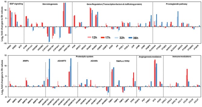
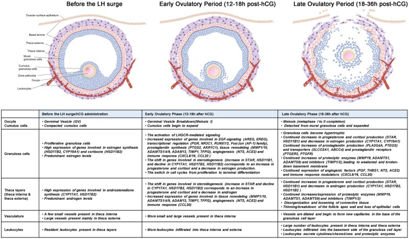
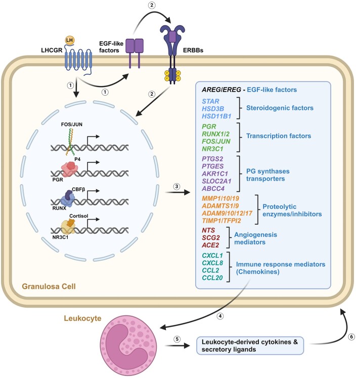
Outcomes: Current findings from studies using in vivo and in vitro models demonstrate that the LH surge or hCG administration increases the expression of ovulatory mediators, including EGF-like factors, steroids, transcription factors, prostaglandins, proteolytic systems, and other autocrine and paracrine factors, similar to those observed in other animal models such as rodents, ruminants, and monkeys. However, the specific ovulatory factors induced, their expression pattern, and their regulatory mechanisms vary among different species. These species-specific differences stress the necessity of utilizing human samples to delineate the mechanisms underlying the ovulatory process in women.

Wider implications: The data from human ovulation in vivo and in vitro models have begun to fill the gaps in our understanding of the ovulatory process in women. Further efforts are needed to discover novel ovulatory factors. One approach to address these gaps is to improve existing in vitro models to more closely mimic in vivo ovulatory conditions in humans. This is critically important as the knowledge obtained from these human studies can be translated directly to aid in the diagnosis of ovulation-associated pathological conditions, for the development of more effective treatment to help women with anovulatory infertility or, conversely, to better manage ovulation for contraceptive purposes.

Registration number: N/A.

Keywords: follicle; granulosa cells; infertility; oocyte; ovulation.


Conflict of interest statement

None declared.

Figures

None
Investigations of the ovulatory process in humans have identified specific signaling pathways and factors expressed in follicular cells and leukocytes, and involved in the complex process of ovulation and luteinization. EGF, epidermal growth factors; PGs, prostaglandins, LUFS, luteinized unruptured follicle syndrome; PCOS, polycystic ovarian syndrome. Image created with BioRender.com
Figure 1.
Figure 1.
In vivo sample collection of granulosa cells from women undergoing IVF/ICSI treatment. Individually dosed rFSH or hMG was given, starting on Days 2–3 of the menstrual cycle. From stimulation Days 5–6, GnRH antagonist was administrated daily. When at least three follicles reached 17 mm in diameter, rhCG or GnRH agonist was given to trigger the ovulatory process. Each woman donated the content of two follicles: one before (0 h, T0) or at 12 h (T12), 17 h (T17), or 32 h (T32), and another one at the time of retrieval at 36 h (T36). The T0 follicle aspiration was taken an average of 11.9 h prior to the ovulation trigger. Granulosa cells and follicular fluid were collected and processed for gene expression analysis and hormone measurement. Image created with BioRender.com.
Figure 2.
Figure 2.
In vivo collection of dominant follicles throughout the periovulatory period from regularly cycling women. Menstrual cyclicity and the size of the dominant follicle were monitored by transvaginal ultrasound. When the dominant follicle reached 14–17 mm, rhCG was given to trigger the ovulatory process. Surgery was performed at one of four predetermined phases: (i) preovulatory phase (Pre), prior to the spontaneous LH surge when the follicle was between 14 and 17 mm; (ii) early ovulatory phase (EO), between 12 and <18 h after hCG; (iii) late ovulatory phase (LO), between 18 and 34 h or less after hCG; or (iv) post-ovulatory phase (PO), between >44–70 h after hCG administration. The dominant follicle was dissected from the ovary and used either for immunohistochemistry or to harvest granulosa cells and thecal tissue for gene expression analysis. Image created with BioRender.com.
Figure 3.
Figure 3.
An image of a dissection of a dominant human follicle of the early ovulatory (EO) stage using laparoscopic scissors and forceps. (A) The dissected whole intact follicle of ∼2 cm in size. (B) Blood vessels can be seen on the surface of the follicle. OC: ovarian cortex; OS: ovarian stroma; FW: follicle wall.
Figure 4.
Figure 4.
Experimental paradigm using in vitro cultures of human granulosa/lutein cells. (A) The in vitro experimental paradigm using human granulosa/lutein cells as described (Al-Alem et al., 2015). Follicles were aspirated at 36 h after ovulation induction with rhCG and cumulus–oocyte complexes were removed from follicular aspirates for fertility treatment. The remaining follicular aspirates were subjected to Percoll-gradient centrifugation to remove blood cells. The isolated luteinizing granulosa cells were plated and cultured for 6 days, with a change of media every 24 h. At the end of acclimation, the cells were washed with the fresh media and treated ± hCG ± selected agents. The cells and culture media were collected after defined hours and used for downstream analyses. (B) Representative microscopic images of human granulosa/lutein cells at the time of plating (Day 0), 6 days after preincubation (Day 6), and 36 h after hCG treatment (1 IU/ml). Magnification of all images, 10×.
Figure 5.
Figure 5.
Temporal changes in gene expression profile in granulosa cells of follicle aspirates collected throughout the ovulatory period. The expression levels of each gene were extracted from the list of differentially regulated genes in the microarray dataset from Poulsen et al. (2020a). Log2-fold change to 0 h levels was calculated using the mean(log2) value of the gene at each time point.
Figure 6.
Figure 6.
HSD11B1 and MMP9 expression in the ovulatory follicle. A dominant follicle was collected at the late ovulatory phase (18–34 h after hCG administration) from a normally cycling woman. Follicle tissue sections were subjected to immunohistochemistry for HSD11B1 and MMP9. The positive staining for HSD11B1 (pink) was localized predominantly to the granulosa cell layer, whereas MMP9-positive cells (purple staining) were localized throughout the theca layer as well as in and outside of blood vessels, suggestive of leukocytes. GC; granulosa cells, TC; theca layer, BV; blood vessel. (*) was noted to mark the same location in serial sections. Bars represent 200 µm.
Figure 7.
Figure 7.
The LH surge/hCG induces temporal and cell-type specific ovulatory changes in the morphology, hormones, and gene expression in human ovulatory follicles. Image created with BioRender.com.
Figure 8.
Figure 8.
Hypothetical model of the ovulatory events occurring in granulosa cells and leukocytes of the dominant follicle in humans. The preovulatory LH surge, upon binding to the LH receptor (LHGCR), triggers the induction of transcription factors and the activation of EGR signaling (1 and 2), leading to an increase in the expression of a diverse array of autocrine/paracrine mediators (3). These mediators include EGF-like factors, steroidogenic enzymes, transcription factors, prostaglandin (PG) synthases and transporters, proteolytic enzymes and their inhibitors, angiogenic mediators, and immune response mediators. Granulosa cell-secreted chemokines play a role in the recruitment of leukocytes (4), which in turn secrete cytokines and secretory ligands (5) that can impact granulosa cell function (6). These inter- and extra-cellular factors coordinate the complex ovulatory process and the subsequent luteinization. The numbers represent the hypothetical order of events occurring in the dominant follicle. P4, progesterone; ERBBs, EGF receptors. Image created with BioRender.com.

References

    1. Adams J, Liu Z, Ren YA, Wun WS, Zhou W, Kenigsberg S, Librach C, Valdes C, Gibbons W, Richards J. Enhanced inflammatory transcriptome in the granulosa cells of women with polycystic ovarian syndrome. J Clin Endocrinol Metab 2016;101:3459–3468. - PMC - PubMed
    1. Akison LK, Robertson SA, Gonzalez MB, Richards JS, Smith CW, Russell DL, Robker RL. Regulation of the ovarian inflammatory response at ovulation by nuclear progesterone receptor. Am J Reprod Immunol 2018;79:e12835. - PubMed
    1. Al-Alem L, Puttabyatappa M, Rosewell K, Brannstrom M, Akin J, Boldt J, Muse K, Curry TE Jr. Chemokine ligand 20: a signal for leukocyte recruitment during human ovulation? Endocrinology 2015;156:3358–3369. - PMC - PubMed
    1. Al-Alem L, Puttabyatappa M, Shrestha K, Choi Y, Rosewell K, Brannstrom M, Akin J, Jo M, Duffy DM, Curry TE. Neurotensin: a neuropeptide induced by hCG in the human and rat ovary during the periovulatory period. Biol Reprod 2021;104:1337–1346. - PMC - PubMed
    1. Andersen CY, Morineau G, Fukuda M, Westergaard LG, Ingerslev HJ, Fiet J, Byskov AG. Assessment of the follicular cortisol:cortisone ratio. Hum Reprod 1999;14:1563–1568. - PubMed