. 2024 Sep 27;10(1):411.
doi: 10.1038/s41420-024-02188-9.

DDB2 expression lights the way for precision radiotherapy response in PDAC cells, with or without olaparib

Affiliations

DDB2 expression lights the way for precision radiotherapy response in PDAC cells, with or without olaparib

Julie Dardare et al. Cell Death Discov. .

Abstract

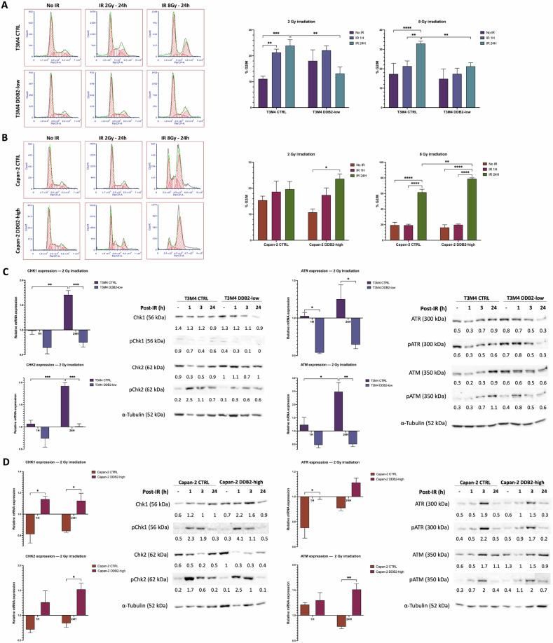
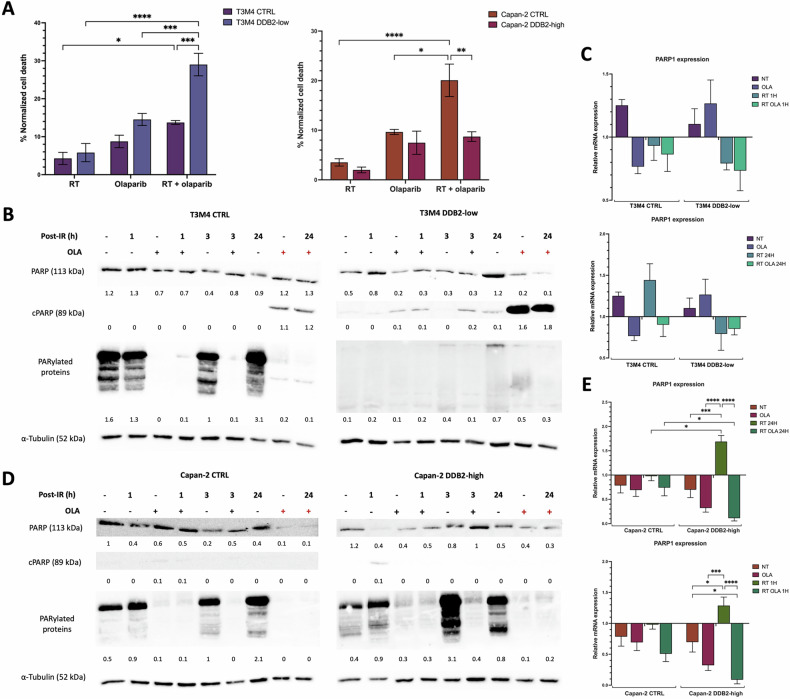
Pancreatic ductal adenocarcinoma (PDAC) is one of the deadliest cancers. Therapeutic options for PDAC are primarily restricted to surgery in the early stages of the disease or chemotherapy in advanced disease. Only a subset of patients with germline defects in BRCA1/2 genes can potentially benefit from personalized therapy, with the PARP inhibitor olaparib serving as a maintenance treatment for metastatic disease. Although the role of radiotherapy in PDAC remains controversial, the use of radiosensitizers offers hope for improving cancer management. Previously, we have shown that damage-specific DNA binding protein 2 (DDB2) is a potential prognostic and predictive biomarker for chemotherapy response in PDAC. In this study, we investigated the function of DDB2 in radiotherapy response, with and without radiosensitization by olaparib in PDAC cells. Our findings demonstrated DDB2 resistance to radiation effects, thereby improving cell survival and enhancing the repair of ionizing radiation-induced DNA double-strand breaks. We observed that DDB2 expression enhances the cell cycle arrest in the G2 phase by phosphorylating Chk1 and Chk2 cell cycle checkpoints. Additionally, we identified a novel link between DDB2 and PARP1 in the context of radiotherapy, which enhances the expression and activity of PARP1. Our findings highlight the potential of low-DDB2 expression to potentiate the radiosensitization effect of olaparib in PDAC cells. Collectively, this study provides novel insights into the impacts of DDB2 in the radiotherapy response in PDAC, enabling its employment as a potential biomarker to predict resistance to radiation. Furthermore, DDB2 represents a significant step forward in precision radiotherapy by widening the scope of patients who can be benefiting from olaparib as a radiosensitizer. Hence, this research has the potential to enrich the limited use of radiotherapy in the care of patients with PDAC.


Conflict of interest statement

The authors declare no competing interests.

Figures

Fig. 1
Fig. 1. DDB2 induces resistance to ionizing radiations in PDAC cells.
A The sensitivity of T3M4 CTRL and T3M4 DDB2-low cells to ionizing radiation was determined by clonogenic formation assay. The survival fraction was determined in relation to the number of colonies obtained in untreated cells. The data from three independent experiments are expressed as the mean ± SEM, *p < 0.05 and ***p < 0.001 (Student’s unpaired t-test). B, C The induction of double-strand breaks was analyzed by γ-H2AX foci labeling. The nucleus was counterstained with DAPI (x40). The mean number of γ-H2AX foci in 50 nuclei was determined at 10 min, 1 h, and 24 h after 2 Gy irradiation. The data from three independent experiments are expressed as the mean ± SEM, *p < 0.05, **p < 0.01, ***p < 0.001, and ****p < 0.0001 (ANOVA).
Fig. 2
Fig. 2. DDB2 affects G2/M arrest through increased expression and phosphorylation of cell cycle checkpoints.
The cell cycle distribution was determined following exposure to 2 Gy and 8 Gy ionizing irradiation (IR) in T3M4 cells (A) and Capan-2 cells (B). The data from three independent experiments are expressed as the mean ± SEM, *p < 0.05, **p < 0.01, ***p < 0.001, and ****p < 0.0001 (ANOVA). The transcriptional and protein expression levels of the cell cycle checkpoint proteins Chk1 and Chk2 and their upstream effectors ATR and ATM were analyzed for T3M4 cells (C) and Capan-2 cells (D). The transcriptional level expression following 1 h and 24 h of exposure to 2 Gy IR were determined by RT-qPCR. β-actin was used as a housekeeping gene. The results were normalized by the expression of the genes of interest in untreated cells. The data from three independent experiments are expressed as mean ± SEM, *p < 0.05, **p < 0.01, and ***p < 0.001 (ANOVA). Protein expression and phosphorylation were studied by western blot analysis following 1 h, 3 h, and 24 h exposure to 2 Gy IR. α-tubulin was used as a loading control.
Fig. 3
Fig. 3. DDB2 impairs the radiosensitizing olaparib effect via PARP1 activity.
A Cell death was monitored by flow cytometry with annexin V and propidium iodide labeling 24 h after 2 Gy radiotherapy alone or 2 Gy radiotherapy with olaparib pretreatment. B, D PARP, cleaved PARP (cPARP), and PARylated proteins expression were studied by western blot after 1 h, 3 h, and 24 h exposure to 2 Gy IR with or without olaparib pretreatment. Olaparib was administered for either 24 h (black +) or 48 h (red +) before. α-tubulin was used as a loading control. C, E PARP1 expression was analyzed by RT-qPCR in different conditions: no treatment (NT), after olaparib treatment (OLA), after radiotherapy treatment (RT), or after radiotherapy with olaparib pretreatment (RT OLA). PARP1 expression was analyzed at 1 h and 24 h post-irradiation. β-actin was used as a housekeeping gene. The expression of untreated cells was normalized to the expression of the corresponding wild-type cell lines, and the expression of the different treatment conditions was normalized to the expression of the corresponding untreated cells. The data from three independent experiments are expressed as the mean ± SEM, *p < 0.05, ***p < 0.001, ****p < 0.0001 (ANOVA).

References

    1. Siegel RL, Miller KD, Wagle NS, Jemal A. Cancer statistics, 2023. CA Cancer J Clin. 2023;73:17–48. - PubMed
    1. Lambert A, Schwarz L, Borbath I, Henry A, Van Laethem JL, Malka D, et al. An update on treatment options for pancreatic adenocarcinoma. Ther Adv Med Oncol. 2019;11:1758835919875568. - PMC - PubMed
    1. Golan T, Hammel P, Reni M, Van Cutsem E, Macarulla T, Hall MJ, et al. Maintenance olaparib for germline BRCA-mutated metastatic pancreatic cancer. N Engl J Med. 2019;25:317–27. - PMC - PubMed
    1. Liao WC, Chien KL, Lin YL, Wu MS, Lin JT, Wang HP, et al. Adjuvant treatments for resected pancreatic adenocarcinoma: a systematic review and network meta-analysis. Lancet Oncol. 2013;14:1095–103. - PubMed
    1. Kalser MH, Ellenberg SS. Pancreatic cancer. Adjuvant combined radiation and chemotherapy following curative resection. Arch Surg. 1985;120:899–903. - PubMed

LinkOut - more resources