. 2024 Sep 27;15(1):8394.
doi: 10.1038/s41467-024-52803-7.

A basally active cGAS-STING pathway limits SARS-CoV-2 replication in a subset of ACE2 positive airway cell models

Affiliations

A basally active cGAS-STING pathway limits SARS-CoV-2 replication in a subset of ACE2 positive airway cell models

Maritza Puray-Chavez et al. Nat Commun. .

Abstract

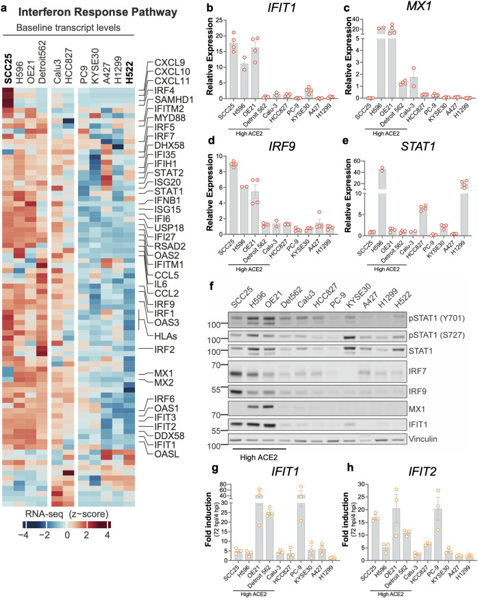
Host factors that define the cellular tropism of SARS-CoV-2 beyond the cognate ACE2 receptor are poorly defined. Here we report that SARS-CoV-2 replication is restricted at a post-entry step in a number of ACE2-positive airway-derived cell lines due to tonic activation of the cGAS-STING pathway mediated by mitochondrial DNA leakage and naturally occurring cGAS and STING variants. Genetic and pharmacological inhibition of the cGAS-STING and type I/III IFN pathways as well as ACE2 overexpression overcome these blocks. SARS-CoV-2 replication in STING knockout cell lines and primary airway cultures induces ISG expression but only in uninfected bystander cells, demonstrating efficient antagonism of the type I/III IFN-pathway in productively infected cells. Pharmacological inhibition of STING in primary airway cells enhances SARS-CoV-2 replication and reduces virus-induced innate immune activation. Together, our study highlights that tonic activation of the cGAS-STING and IFN pathways can impact SARS-CoV-2 cellular tropism in a manner dependent on ACE2 expression levels.


Conflict of interest statement

The authors declare no competing interests.

Figures

Fig. 1
Fig. 1. SARS-CoV-2 replication is blocked at a post-entry stage in a subset of airway-derived cell lines that endogenously express ACE2.
a The indicated airway-derived cell lines were inoculated with WT SARS-CoV-2 (2019-nCoV/USA-WA1/2020) at a MOI: 0.15 i.u./cell. RT-qPCR analysis of cell-associated SARS-CoV-2 N sgRNA (copies/cell) at 4 and 72 hpi is shown. Data show the average of 2 independent experiments, error bars denote the range. b Focus forming assay performed on culture supernatants of the indicated cells infected with SARS-CoV-2 for 72 hpi (from panel a). Data show focus forming units per mL (FFU/mL, log transformed) from two independent experiments, error bars show the SEM. c Polyclonal pools of ACE2 knockout (ACE2-KO) and control cell lines transduced with a non-targeting sgRNA (NT) were infected with WT SARS-CoV-2 at an MOI: 2 i.u./cell. RT-qPCR analysis of cell-associated viral RNA (sgRNA N) at 72 hpi is shown from two biological replicates. Data show the mean, error bars show the SEM. d Polyclonal pools of ACE2 KO and non-targeting control (NT) cell lines were infected with WT SARS-CoV-2 at a MOI: 2 i.u./cell. In situ hybridization of incoming viral RNA (green) at 2 hpi was performed as detailed in Methods. Nuclear DAPI staining is shown in blue. e Quantification of data presented in Panel d. Puncta of SARS-CoV-2 RNA was counted in ∼200 cells per cell line from two independent experiments. The average number of RNA puncta per condition is shown in the graph. Source data are provided as a Source Data file.
Fig. 2
Fig. 2. ACE2(high) airway-derived cell lines that are resistant to SARS-CoV-2 replication express high baseline levels of type I IFN pathway genes.
a Hierarchical clustering of IFN response pathway RNA expression levels downloaded from CCLE. The heatmap displays z-scored log2(TPM) values. b–e RT-qPCR analysis of representative ISGs, including IFIT1, MX1, IRF9 and STAT1 in uninfected cells. Expression levels are normalized relative to Calu-3 cells (set to 1). Data show the mean from n = 2 (H596 and Calu-3), or 4 (other cells) independent biological replicates, error bars denote SEM. f Immunoblot showing pSTAT1 (Y701 and S727), STAT1, IRF7, IRF9, MX1, IFIT1, and Vinculin levels across airway-derived cell lines (not infected). Blots are representative of two independent experiments. g, h Airway cell lines were infected with SARS-CoV-2 at an MOI of 2 i.u./cell, and expression of IFIT1 and IFIT2 was analyzed by RT-qPCR at 4 and 72 hpi. Y-axes indicate ISG fold induction at 72 hpi relative to 4hpi. Data show the mean from n = 3 independent replicates, error bars show the SEM. Source data are provided as a Source Data file.
Fig. 3
Fig. 3. Basally active type I IFN pathway limits SARS-CoV-2 replication in a subset of ACE2(high) cells.
a, b SCC25, H596, OE21, Detroit 562, and Calu-3 cells were pretreated with 1 μM ruxolitinib for 24 h. IFIT1 (a) and MX1 (b) expression were analyzed by RT-qPCR, and expression levels are shown relative to untreated Calu-3 cells (set to 1). Data show the mean from n = 2 (H596 and Calu-3) and 4 (other cells) independent biological replicates, error bars show the SEM. c SCC25, H596, OE21, and Detroit 562 cells were pretreated with DMSO (mock) or 1 μM ruxolitinib for 24 h and infected with SARS-CoV-2 at a MOI of 2 i.u./cell in the presence of DMSO or 1 μM ruxolitinib. RT-qPCR analysis shows cell-associated viral RNA levels at 72 hpi. Data show the mean from n = 4 independent replicates, error bars show the SEM. d–g Polyclonal populations of SCC25 cells knocked out (KO) for IFNAR (d) or STAT1 (e) using two independent sgRNAs or transduced with a non-targeting (NT) control and SCC25 cells transfected with 3 separate siRNAs targeting STAT2 (f), IRF9 (g), or a non-targeting control (NT) were infected with SARS-CoV-2 at a MOI of 2 i.u./cell. Cell-associated viral RNA was analyzed by RT-qPCR at 72 hpi. Y-axes indicate the relative expression ratio between knockdown (KD) or knockout (KO) and the respective NT samples. Data display the mean of n = 5 (d, e) and n = 6 (f, g) independent replicates, error bars denote the SEM. Source data are provided as a Source Data file.
Fig. 4
Fig. 4. A constitutively active cGAS-STING pathway limits SARS-CoV-2 replication in SCC25 cells.
a SCC25 cells transfected with siRNAs or a non-targeting control (NT) were infected with SARS-CoV-2 at an MOI of 2 i.u./cell for 72 h. Data show the ratio of cell-associated viral RNA between knockdown (KD) and NT samples. Data display the mean of 2 (NLRP1-3, NLRP3-1, NLRP3-2, NLRC5-1, NLRC5-2) or 4 (other targets) biological replicates, with error bars representing SEM. b SCC25 cGAS, STING, TBK1 and IRF3 KO cells or NT controls were infected with SARS-CoV-2 at MOI: 2 i.u./cell for 72 h. Cell-associated viral N RNA from 5 replicates is shown, and error bars show the SEM. p < 0.0001 significance (compared to NT-1) was assessed by a one-way ANOVA with Dunnett correction. c Immunoblots (representative of n = 2) show SARS-CoV-2 N, cGAS, STING, TBK1, IRF3 and β-actin expression levels in SCC25 cells from panel b. d SCC25 STING KO cells were infected with SARS-CoV-2-mNG at a MOI:2 i.u./cell. NG-positive cells were enumerated by flow cytometry at 24, 48, and 72 hpi and represented as a percentage of the total population. Data show the mean of n = 5 independent experiments, error bars denote the SEM. e SCC25 STING KO and NT control cells were pretreated for 24 h and infected (MOI:2 i.u./cell) in the absence (DMSO) and presence of 1 μM ruxolitinib with SARS-CoV-2-mNG. NG-positive cells were enumerated by flow cytometry at 72 hpi and represented as a percentage of the total population. Data show the mean of n = 4 independent experiments, error bars denote the SEM. P-values are assessed by two-way ANOVA and Šidák multiple test correction. f Cells ectopically expressing ACE2 were infected at a MOI: 0.1 i.u./cell with SARS-CoV-2 WT. Cell-associated SARS-CoV-2 RNA at 2 and 24 hpi is shown from n = 2 replicates. Data show the mean, error bars denote SEM. g, h SCC25 (g) and OE21 (h) cells were transfected with 2 distinct siRNAs or an NT control, infected with SARS-CoV-2 and analyzed as in panel a). Data display the mean of n = 3 biological replicates, with error bars representing SEM. Source data are provided as a Source Data file.
Fig. 5
Fig. 5. Genetic ablation and chemical inhibition of STING reduces basal ISG levels and enhances SARS-CoV-2 replication.
a WT and STING KO SCC25, H596, OE21, and Detroit 562 cells were subjected to RNA-seq analysis. Heatmap shows differentially expressed genes (absolute log2 fold change of > 2 and an adjusted q value of < 0.05 from n = 4 independent replicates for each cell type) in the IFN and inflammatory pathways in any given cell line between the WT vs. STING KO variant. b–g RT-qPCR analysis of IFIT2, MX1 and IFIT3 in uninfected SCC25, H596, OE21 and Detroit 562 cells treated with increasing concentrations of SN-011 (bd) and H-151 (eg) for 48 h. Data show the mean from n = 2 independent biological replicates, error bars show SEM (h, i) WT and STING KO SCC25 and STING KO cells were pre-treated with 20 μM SN-011 (h) or H-151 (i) for 24 h  were infected with WT SARS-CoV-2 at MOI: 1 i.u./cell for 72 h. Data show the copies of cell-associated SARS-CoV-2 N RNA in compound-treated cells normalized relative to mock-treated samples. Data show the mean from n = 3 independent biological replicates, error bars show SEM. Significance was assessed by multiple unpaired two-tailed t tests and p-values provided in the figure. Source data are provided as a Source Data file.
Fig. 6
Fig. 6. Cell culture supernatants but not cell lysates from SCC25, H596, and OE21 cells trigger innate immune activation.
a, b MX1 expression was analyzed by RT-qPCR in THP-1 cells treated with cell culture supernatants from (a) or co-cultured with (b) the indicated cell line panel as explained in Methods. MX1 expression is normalized relative to mock-treated cells. THP-1 cells treated with 1000 units/ml IFN-α serve as the positive control. Data show the mean induction from n = 3 independent biological replicates, error bars display the SEM. p-values were calculated by multiple unpaired t-tests with Welch’s correction. c, d THP-1 or THP-1 STING KO cells were treated with post-nuclear supernatants (PNS) from the indicated cells, 10 μg/ml 2’3’-cGAMP or 10 μg/ml 2’2’-cGAMP for 24 h, as explained in Methods. IFN-β expression was analyzed by RT-qPCR and normalized relative to 2’2’-cGAMP-treated samples. Data show the mean from n = 2 independent experiments, error bars show the SEM. Source data are provided as a Source Data file.
Fig. 7
Fig. 7. SARS-CoV-2 antagonizes the IFN pathway but not IFN synthesis or inflammatory pathways in infected cells.
a Relative expression of ISGs including ISG15, IFIT1, IFIT2, IFIT3, and IRF7 in SCC25 cGAS KO, STING KO or NT control cells infected with SARS-CoV-2 at a MOI of 2 i.u./cell. Cells were collected at 72 hpi, and ISG expression was analyzed by RT-qPCR. Data show the fold induction of ISGs in infected over uninfected cells from n = 3 biological replicates, errors bars show the SEM. b, c SCC25 STING KO or NT control cells were infected with SARS-CoV-2 at an MOI of 2 i.u./cell. Immunofluorescence detection for IFIT3 expression (in red) and SARS-CoV-2 nucleocapsid (N) viral protein (in green), and cellular nuclei (DAPI, in blue) at 72 hpi from a representative experiment (n = 2) are shown. Images were collected with an epifluorescence microscope, 4X objective (b), or Zeiss LSM 880 Airyscan confocal microscope equipped with a × 63/1.4 objective (c) as detailed in Methods. Scale bars = 250 μm (b) or 10 μm (c). d, e SCC25 STING KO cells were infected with SARS-CoV-2-mNG at MOI:1 i.u./cell, and cells were single-cell sorted at 72 hpi to isolate infected (mNG positive) and bystander cells as detailed in Methods. Expression of the indicated ISGs (d) and inflammatory genes (e) were analyzed by RT-qPCR in these sorted cell populations. Data show the mean from two independent replicates, error bars show the SEM. Source data are provided as a Source Data file.
Fig. 8
Fig. 8. Analysis of mechanisms that underlie tonic cGAS-STING activation in the ISG(high) cell lines.
a Polyclonal populations of SCC25, H596, OE21, and Detroit 562 cells knocked out (KO) for TREX1 using two separate sgRNAs or wild-type (WT) controls were analyzed for IFIT2 expression by RT-qPCR. Data show the relative expression of IFIT2 in KO cells compared to parental controls (set to 1) from n = 3 (OE21) or 6 (SCC25, H596, Detroit 562) replicates, error bars show the SEM. b cGAS and STING variants present in the indicated cell lines derived from RNA-seq data. c, d SCC25, H596, OE21, and Detroit 562 cells grown in the presence of 10 μM VBIT-4 for 4 days (c) or 500 μM AZT for 3 days (d). IFIT1, IFIT2, and IRF7 expression was analyzed by RT-qPCR and normalized relative to mock-treated controls (set to 1). Data show the mean from n = 3 replicates, error bars show the SEM. e Following subcellular fractionation (see “Methods”), levels of the indicated HERV and LINE DNA elements were assessed by qPCR in the cytosol and nucleus. Data show the relative levels of each element in the cytosol relative to the nuclear extract in each cell type (n = 2, error bars show SEM). Source data are provided as a Source Data file.
Fig. 9
Fig. 9. Analysis of ISG and STING expression in SARS-CoV-2-infected primary human airway epithelial cells (hTECs) grown at ALI.
ae hTECs from five donors grown at ALI were infected with SARS-CoV-2-mNG at an MOI of 2 i.u./cell and fixed at 72 hpi. Immunofluorescence detection for IFIT2 expression (in red), ISG15, mNG (in green), and cellular nuclei (DAPI, in blue) is shown (a). Images were analyzed by confocal microscopy with a × 40/1.4 objective. Scale bars = 20 μm, (representative of n = 2). Pearson’s correlation (b, d) and Mander’s overlap (c, e) coefficient were derived from imaging experiments. Colocalization analysis of Z stacks was performed using the Colocalization module of Volocity from 3 to 8 separate Z-stacks. Individual numbers plotted display the corresponding values from each Z-stack for each Donor, error bars show the SEM. f, g hTECs infected as in panel A were stained for STING (in red), SARS-CoV-2 N (in green), and cellular nuclei (DAPI, in blue). Images (f) were analyzed by confocal microscopy with a × 20 objective. Scale bars = 50 μm, (representative of n = 2). Colocalization analysis of Z stacks was performed as above, and Pearson’s correlation coefficient derived from 3 to 6 separate Z-stacks was plotted, error bars show SEM (g). h, i hTECs from three donors were pretreated with 20 μM SN-011 or H-151 for 48 h (both apical and basal chambers) and infected with SARS-CoV-2 mNG as above in the presence of compounds in the basal chamber. Virus release in the ALI chamber (h) and expression levels of the indicated ISGs (i) were analyzed by RT-qPCR. Data in (h) show the copies of cell-free SARS-CoV-2 N RNA in culture supernatants of compound-treated cells normalized relative to mock-treated samples. Data show the mean from n = 2 independent biological replicates, error bars show SEM. Source data are provided as a Source Data file.
Fig. 10
Fig. 10. Expression profiles of cGAS, STING, and representative ISGs in the healthy human airways.
Normalized counts and cell type metadata were downloaded from a publicly available single-cell study. Detection frequency represents the percentage of cells with > 0 counts for the respective gene. Bar plots display the average normalized expression for a gene within a cell type. For the gene set representing the interferon alpha Gene Ontology term, normalized expression levels were summed for all genes in the set prior to computing detection frequencies and average expression.

Update of

References

    1. Chen, N. et al. Viral strategies to antagonize the host antiviral innate immunity: an indispensable research direction for emerging virus-host interactions. Emerg. Microbes Infect.13, 2341144 (2024). - PMC - PubMed
    1. Le Pen, J. & Rice, C. M. The antiviral state of the cell: lessons from SARS-CoV-2. Curr. Opin. Immunol.87, 102426 (2024). - PMC - PubMed
    1. Sievers, B. L., Cheng, M. T. K., Csiba, K., Meng, B. & Gupta, R. K. SARS-CoV-2 and innate immunity: the good, the bad, and the “goldilocks. Cell Mol. Immunol.21, 171–183 (2024). - PMC - PubMed
    1. Walls, A. C. et al. Structure, Function, and Antigenicity of the SARS-CoV-2 Spike Glycoprotein. Cell181, 281–292.e286 (2020). - PMC - PubMed
    1. Letko, M., Marzi, A. & Munster, V. Functional assessment of cell entry and receptor usage for SARS-CoV-2 and other lineage B betacoronaviruses. Nat. Microbiol.5, 562–569 (2020). - PMC - PubMed

Publication types

Associated data