. 2024 Sep 27;15(1):8299.
doi: 10.1038/s41467-024-52584-z.

Cryo-EM structure of human class C orphan GPCR GPR179 involved in visual processing

Affiliations

Cryo-EM structure of human class C orphan GPCR GPR179 involved in visual processing

Yaejin Yun et al. Nat Commun. .

Erratum in

Abstract

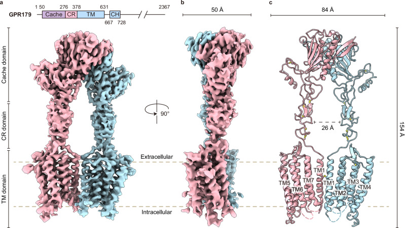
GPR179, an orphan class C GPCR, is expressed at the dendritic tips of ON-bipolar cells in the retina. It plays a pivotal role in the initial synaptic transmission of visual signals from photoreceptors, and its deficiency is known to be the cause of complete congenital stationary night blindness. Here, we present the cryo-electron microscopy structure of human GPR179. Notably, the transmembrane domain (TMD) of GPR179 forms a homodimer through the TM1/7 interface with a single inter-protomer disulfide bond, adopting a noncanonical dimerization mode. Furthermore, the TMD dimer exhibits architecture well-suited for the highly curved membrane of the dendritic tip and distinct from the flat membrane arrangement observed in other class C GPCR dimers. Our structure reveals unique structural features of GPR179 TMD, setting it apart from other class C GPCRs. These findings provide a foundation for understanding signal transduction through GPR179 in visual processing and offers insights into the underlying causes of ocular diseases.


Conflict of interest statement

The authors declare no competing interests.

Figures

Fig. 1
Fig. 1. Cryo-EM structure of human GPR179.
a Schematic diagram showing the subdomain composition in GPR179 and a cryo-EM map of GPR179 (front view). The Cache domain, cysteine-rich domain (CR domain), transmembrane domain (TM domain), and cytoplasmic helices (CH) are colored in violet, light pink, and light blue, respectively. Chains A and B are represented as light pink and light blue, respectively. b Cryo-EM map of GPR179 (side view). c Model of GPR179 (front view) colored the same as in (a). Transmembrane helices are denoted as TM.
Fig. 2
Fig. 2. Noncanonical dimeric arrangement of TMD and surrounding membrane curvature for GPR179.
a The overall structure of the TM domain (TMD) homodimer. Chains A and B are depicted as light pink and light blue, respectively, with disulfide bonds shown as yellow sticks. The black arrows indicate extracellular loop (ECL). b An overview of the GPR179 TMD from both the top and bottom views. The red line indicates the distance between the two closest TM from two protomers. The TMD dimer interface is enclosed in a gray box (front view), and detailed interactions at the TMD dimer interface (TM1/7) are illustrated. Disulfide bonds are shown as yellow sticks. c A bottom view of the GPR179 TMD arrangement compared to mGlyR (PDB code 7EWL, gray), mGluR5 (PDB code 6N52, orange), and GABAB (PDB code 7C7S, green) in their inactive states. The chain A protomers of GPR179, mGlyR, mGluR5, and GABAB are superimposed. d A bottom view of the GPR179 TMD arrangement compared to mGluR5 (PDB code 6N51, dark orange) and GABAB (PDB code 7C7Q, light green) in their active states. The chain A protomers of GPR179, mGlyR, mGluR5, and GABAB are superimposed. e Membrane curvature for GPR179. A comparison of the structures of GPR179 with other class C GPCRs, where membrane-embedded regions were computed using the Orientations of Proteins in Membranes server (https://opm.phar.umich.edu/ppm_server3): GPR179, mGlyR (PDB code 7SHE), mGluR5 (PDB code 6N52), and GABAB (PDB code 7C7S). The radius of intrinsic curvature (R) of spatial positions in spherical membranes was calculated by PPM 3.0. The membrane is indicated by purple spheres.
Fig. 3
Fig. 3. Dissection of GPR179 TMD protomer.
a Comparison of TM3 and TM4 helix lengths among class C GPCRs. Chain A protomers of GPR179 (light pink), mGlyR (gray, PDB code 7EWL), and GABAB (green, PDB code 7C7S) are superimposed. b The interactions in the ECL region including extracellular domain (ECD)-transmembrane helix 1 (TM1) loop. Yellow sticks represent disulfide bonds, while residues participating in hydrophobic and polar interactions (black dashed lines) are shown. c Overall structure of the GPR179 TMD protomer, divided into the upper, middle, and lower parts from top to bottom. d Electrostatic interactions within the upper part of the TMD for GPR179 and mGlyR (PDB code 7EWL) (top view). e The polar interactions among TM3, TM5, TM6, and TM7 in the middle part for GPR179. f The ionic and polar interaction network in the lower part (bottom view), including the intracellular loop (ICL). Notably, K6307.51, belonging to the conserved FxPKxx motif found in class C GPCRs, interacts with the “ionic lock”.
Fig. 4
Fig. 4. Structural comparison of ECDs of GPR179 and mGlyR.
a Overall structure of the GPR179 ECD. The six pairs of disulfide bonds are shown as yellow sticks. b Top view of the Cache domain dimer interface for GPR179. c Structural comparison of the Cache domain dimer interface between GPR179 and mGlyR (PDB code 7EWL). d The ECD dimer interface in Cache domain. Hydrophobic and electrostatic interactions were depicted between protomers of GPR179 or mGlyR (PDB code 7EWL). e Distinct conformation of the GPR179 homodimer compared with mGlyR. The distance between αI helices in GPR179 (based on Pro93) is 7 Å, whereas the distance is 14 Å in mGlyR (based on Pro120). The distance between CRDs in GPR179 is 25 Å (based on Pro321), while it is 42 Å in mGlyR (based on Ala 356). The loop connecting the ECD and TM1 is indicated by a black arrow.
Fig. 5
Fig. 5. Missense mutation sites associated with cCSNB in GPR179.
The structure of GPR179 with cCSNB mutations. Disease-causing mutation sites identified in cCSNB are highlighted as black circles and magnified views are shown in black boxes. The mutated residues (His126, Cys220, Asp455, and Tyr603) in cCSNB are shown in violet sticks.
Fig. 6
Fig. 6. The putative ligand-binding site.
a Schematic representation of the BRET assay performed to monitor the GAP activity of RGS7-Gβ5 and GPR179. The panel was created with BioRender.com released under a Creative Commons Attribution-NonCommercial-NoDerivs 4.0 International license https://creativecommons.org/licenses/by-nc-nd/4.0/deed.en. b GAP assay measuring kinetics of Gαo deactivation catalyzed by GPR179 in the absence or presence of glycine (left panel). Quantification of GAP activity measured in the right panel (n = 9 independent experiments). c Overall structural organization of Cache domains of mGlyR and GPR179 with putative ligand-binding pockets. Surface filing model show distribution of electrostatic potentials in red and blue colors (–5 to +5 kT/e; red, negative; blue, positive; PyMol using APBS tools). Side chains of key conserved residues are shown as balls. Analysis of putative binding pocket of receptors show that GPR179 has acidic while mGlyR has amphipathic pocket. d Quantification of the GAP activity of GPR179-identified mutants (D126H, Y220C, G455D, H603Y, and C390A) (n = 12 independent experiments) **p value pcDNA3.1 (Control) vs G455D = 0.009 < 0.01. e BRET parameters of the basal state (9.9 sec) before initiating or terminating Gαo signaling of GPR179 mutants (n = 12 independent experiments) *p value WT vs H603Y = 0.0489 < 0.05. *p value WT vs PGL-AAA = 0.0436 < 0.05. Data represent mean ± SEM. **p < 0.01, *p < 0.05, ns (not significant) = p > 0.05, one-way ANOVA.

References

    1. Schiller, P. H., Sandell, J. H. & Maunsell, J. H. Functions of the ON and OFF channels of the visual system. Nature322, 824–825 (1986). - PubMed
    1. Ray, T. A. et al. GPR179 is required for high sensitivity of the mGluR6 signaling cascade in depolarizing bipolar cells. J. Neurosci.34, 6334–6343 (2014). - PMC - PubMed
    1. Werblin, F. S. & Dowling, J. E. Organization of the retina of the mudpuppy, Necturus maculosus. II. Intracellular recording. J. Neurophysiol.32, 339–355 (1969). - PubMed
    1. Nelson, R. & Connaughton, V. In: Webvision: The Organization of the Retina and Visual System (eds. H. Kolb, E. Fernandez, & R. Nelson) (University of Utah Health Sciences Center Copyright: © 2023 Webvision. 1995). - PubMed
    1. Wilmet, B. et al. Mice lacking Gpr179 with complete congenital stationary night blindness are a good model for myopia. Int. J. Mol. Sci.24, 219 (2022) - PMC - PubMed

Publication types

Substances

Supplementary concepts

LinkOut - more resources