. 2024 Sep 27;14(1):22341.
doi: 10.1038/s41598-024-73386-9.

Psychiatric phenotype in neurodevelopmental myoclonus-dystonia is underpinned by abnormality of cerebellar modulation on the cerebral cortex

Affiliations

Psychiatric phenotype in neurodevelopmental myoclonus-dystonia is underpinned by abnormality of cerebellar modulation on the cerebral cortex

Clément Tarrano et al. Sci Rep. .

Abstract

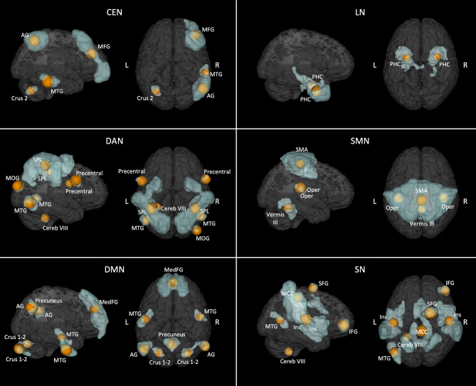
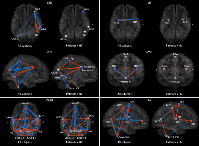
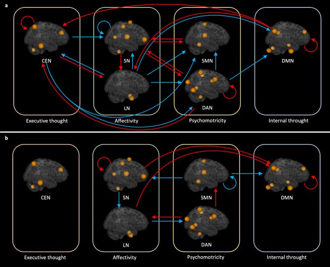
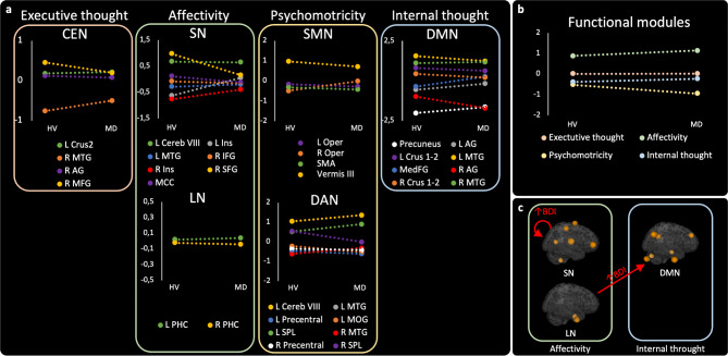
Psychiatric symptoms are common in neurodevelopmental movement disorders, including some types of dystonia. However, research has mainly focused on motor manifestations and underlying circuits. Myoclonus-dystonia is a rare and homogeneous neurodevelopmental condition serving as an illustrative paradigm of childhood-onset dystonias, associated with psychiatric symptoms. Here, we assessed the prevalence of psychiatric disorders and the severity of depressive symptoms in patients with myoclonus-dystonia and healthy volunteers (HV). Using resting-state functional neuroimaging, we compared the effective connectivity within and among non-motor and motor brain networks between patients and HV. We further explored the hierarchical organization of these networks and examined the relationship between their connectivity and the depressive symptoms. Comparing 19 patients to 25 HV, we found a higher prevalence of anxiety disorders and more depressive symptoms in the patient group. Patients exhibited abnormal modulation of the cerebellum on the cerebral cortex in the sensorimotor, dorsal attention, salience, and default mode networks. Moreover, the salience network activity was directed by the cerebellum in patients and was related to depressive symptoms. Altogether, our findings highlight the role of the cerebellar drive on both motor and non-motor cortical areas in this disorder, suggesting cerebellar involvement in the complex phenotype of such neurodevelopmental movement disorders.

Keywords: Cerebellum; Dystonia; MRI; Myoclonus; Network.


Conflict of interest statement

C. Galléa, C. Tranchant, B. Beranger, V. Brochard, C. Atkinson-Clement, L. Defebvre, P. Krystkowiak, J-L. Houeto, J.C. Corvol, E. Apartis, D. Martino, (A) Degardin, J-M Pedespan, M. Vidailhet, Y. Worbe have no disclosures in relation to this work.C. Tarrano received a PhD grant from the “Fondation pour la Recherche Médicale”. C. Delorme has received travel funding from Merz Pharma, Abbvie, and received honoraria from Merz Pharma. (B) Degos received honoraria for acting as a member of Scientific Advisory Boards/Consultancy for Merz Pharma, speaker honoraria from Merz Pharma, Abbvie and Ipsen, and travel support for congresses from Merz Pharma. S. Thobois received honorarium from Abbvie and Merz. E. McGovern has received speaking honoraria from AbbVie, Dutch MDS symposium, served on an advisory board for AbbVie and received research grants from the STAR MD and the RCSI Richard Steeven’s Scholarship. D Grabli served on scientific advisory boards for AbbVie, received speech honoraria from Abbvie, Merz and received travel funding from Abbvie and Merz. E.Roze. received honorarium for participating in an advisory board from Merz-Pharma, received research support from Merz-Pharma, Ipsen, AMADYS, Agence Nationale de la Recherche, Societé Française de Médecine Esthétique, Dystonia Medical Reasearch Foundation.

Figures

Fig. 1
Fig. 1
Results of the Independent Component analysis. In blue, the networks of interest identified by the Independent Component analysis. In orange, the 8 mm spherical nodes (i.e., those used for the effective connectivity analysis) derived from the parcellation of each network. DAN = Dorsal attention network; SMN = Sensorimotor network; LN = Limbic network; SN = Salience network; DMN = Default mode network; CEN = Central executive network; SMA = Supplementary motor area; Oper = Rolandic operculum; SPL = Superior parietal lobule; MTG = Middle temporal gyrus; MOG = Middle occipital gyrus; IFG = Inferior frontal gyrus; SFG = Superior frontal gyrus; MCC = Middle cingulate cortex; PHC = Parahippoccampal cortex; Cereb = Cerebellum; Ins = Insula; MFG = Middle frontal gyrus; AG = Angular gyrus; MedFG = extended Median frontal region; L = Left; R = Right.
Fig. 2
Fig. 2
Results of the within-network effective connectivity analysis. For each network, in the left image red and blue represent respectively excitatory and inhibitory connections across all participants (i.e. patients and controls), whereas in the right image red and blue represent respectively increased excitation (or decreased inhibition) and increased inhibition (or decreased excitation) in patients compared to healthy volunteers (HV). Only the connections with a posterior probability > 0.99 are displayed. DAN = Dorsal attention network; SMN = Sensorimotor network; LN = Limbic network; SN = Salience network; DMN = Default mode network; CEN = Central executive network; SMA = Supplementary motor area; Oper = Rolandic operculum; SPL = Superior parietal lobule; MTG = Middle temporal gyrus; MOG = Middle occipital gyrus; IFG = Inferior frontal gyrus; SFG = Superior frontal gyrus; MCC = Middle cingulate cortex; PHC = Parahippoccampal cortex; Cereb = Cerebellum; Ins = Insula; MFG = Middle frontal gyrus; AG = Angular gyrus; MedFG = extended Median frontal region; L = Left; R = Right.
Fig. 3
Fig. 3
Results of the between-network effective connectivity analysis.Red and blue arrows represent, respectively, excitatory and inhibitory connections between networks across all participants. b Red and blue arrows represent, respectively, increased excitation (or decreased inhibition) and increased inhibition (or decreased excitation) in patients with myoclonus-dystonia. Only the connections with a posterior probability > 0.99 are displayed. DAN = Dorsal attention network ; SMN = Sensorimotor network ; LN = Limbic network ; SN = Salience network ; DMN = Default mode network ; CEN = Central executive network.
Fig. 4
Fig. 4
Hierarchical organization of networks and relationships between change of connectivity and BDI scores in patients. a Comparison of the hierarchical organization within each network between the groups of healthy volunteers (HV) and patients (MD). Nodes at the top act as drivers, while nodes at the bottom serve as sinks. b Comparison of the hierarchical organization of functional units between the groups of healthy volunteers and patients. c, results of the analysis of the DCM PEB model with the Beck depression inventory as covariate of interest in the group of patients. Only connections that differed between groups were considered. DAN = Dorsal attention network; SMN = Sensorimotor network; LN = Limbic network; SN = Salience network; DMN = Default mode network; CEN = Central executive network; SMA = Supplementary motor area; Oper = Rolandic operculum; SPL = Superior parietal lobule; MTG = Middle temporal gyrus; MOG = Middle occipital gyrus; IFG = Inferior frontal gyrus; SFG = Superior frontal gyrus; MCC = Middle cingulate cortex; PHC = Parahippoccampal cortex; Cereb = Cerebellum; Ins = Insula; MFG = Middle frontal gyrus; AG = Angular gyrus; MedFG = extended Median frontal region; L = Left; R = Right.

References

    1. Ben-Pazi, H., Jaworowski, S. & Shalev, R. S. Cognitive and psychiatric phenotypes of movement disorders in children: a systematic review: review. Dev. Med. Child. Neurol.53, 1077–1084 (2011). - PubMed
    1. Kuyper, D. J., Parra, V., Aerts, S., Okun, M. S. & Kluger, B. M. Nonmotor manifestations of dystonia: a systematic review. Mov. Disord. 26, 1206–1217 (2011). - PMC - PubMed
    1. Balint, B. et al. Dystonia. Nat. Rev. Dis. Primer. 4, 25 (2018). - PubMed
    1. Roze, E., Lang, A. E. & Vidailhet, M. Myoclonus-dystonia: classification, phenomenology, pathogenesis, and treatment. Curr. Opin. Neurol.31, 484–490 (2018). - PubMed
    1. Peall, K. J. et al. Psychiatric disorders, myoclonus dystonia and SGCE: an international study. Ann. Clin. Transl Neurol. 3, 4–11 (2016). - PMC - PubMed

Supplementary concepts

LinkOut - more resources