Review
. 2024 Nov;27(11):2056-2072.
doi: 10.1038/s41593-024-01701-8. Epub 2024 Sep 27.

Advances and controversies in meningeal biology

Affiliations
Review

Advances and controversies in meningeal biology

Christer Betsholtz et al. Nat Neurosci. 2024 Nov.

Abstract

The dura, arachnoid and pia mater, as the constituent layers of the meninges, along with cerebrospinal fluid in the subarachnoid space and ventricles, are essential protectors of the brain and spinal cord. Complemented by immune cells, blood vessels, lymphatic vessels and nerves, these connective tissue layers have held many secrets that have only recently begun to be revealed. Each meningeal layer is now known to have molecularly distinct types of fibroblasts. Cerebrospinal fluid clearance through peripheral lymphatics and lymph nodes is well documented, but its routes and flow dynamics are debated. Advances made in meningeal immune functions are also debated. This Review considers the cellular and molecular structure and function of the dura, arachnoid and pia mater in the context of conventional views, recent progress, and what is uncertain or unknown. The hallmarks of meningeal pathophysiology are identified toward developing a more complete understanding of the meninges in health and disease.


Conflict of interest statement

Competing interests

The authors declare no competing interests.

Figures
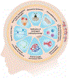
Fig. 1 |
Fig. 1 |. Hallmarks of meningeal pathophysiology.
Illustration of the diverse and distinctive functions of the meninges as they relate to health and disease. The meninges, consisting of the outermost dura, central arachnoid, and innermost pia, are connective tissue layers that enclose cerebrospinal fluid (CSF) in the subarachnoid space (SAS) and ventricles and serve as mechanical protectors of the CNS. The dura contains lymphatic vessels and is innervated by nociceptive sensory axons, derived mainly from neurons of the trigeminal ganglion, that contribute to the symptoms and pathophysiology of migraine. The arachnoid barrier layer of the meninges has tight intercellular junctions that create a restrictive permeability barrier. Other regions are sites for regulated immune surveillance by a diverse repertoire of immune cells. Bidirectional communication for immune cell migration between skull bone marrow and dura through veins in bone channels has been proposed and is currently being studied. The SAS serves as a reservoir for CSF circulation and clearance through mechanisms involving dural lymphatics that drain to lymph nodes in the neck and elsewhere. The SAS also contains arteries that supply the brain and veins that drain into dural venous sinuses. These diverse meningeal functions can be compromised by traumatic head injury, hemorrhage, meningitis, or meningioma, and can also be impaired in neuroinflammatory and neurogenerative conditions and by metastases from tumors in other organs.
Fig. 2 |
Fig. 2 |. Composition of meningeal membranes, barriers, extracellular matrix, and sites of hemorrhage.
The arachnoid and pial membranes consist of five transcriptionally distinct types of fibroblasts: dural border cells (brown); arachnoid barrier cells (pink); two types of inner arachnoid cells (blue and turquoise); and pial and perivascular fibroblasts (green). Also shown are dural fibroblasts (grey), dural immune cells (blue), dural nerves (brown), collagen fibrils (black), basement membrane (violet), blood vessels and lymphatics (yellow). Distinctive features of the meninges include: a. Dural fibroblasts are dispersed among dense bundles of collagen fibrils that form most of the dural volume. b. Dural border cell layers have little extracellular matrix and are sites of potential separation and subdural space formation. c. Arachnoid barrier cells in two overlapping layers are connected by adherens junctions and sealed by tight junctions and tricellular junctions. d. Inner arachnoid cells form two discontinuous layers, one Prox1+ and the other Sidt1+, that are structurally supported by basement membrane, collagen fibrils, and other extracellular matrix components. e. The subarachnoid space is spanned by trabeculae from the inner arachnoid to the pia that vary in size, density, and collagen content in different CNS regions and in different species. f. The pial membrane is a largely continuous cell monolayer with scattered intercellular openings (stomata). g. Pial fibroblasts are transcriptionally closely related to and merge with fibroblasts around blood vessels in the subarachnoid space and penetrating the brain parenchyma. h. The subpial space contains a meshwork of collagen fibrils that provide structural support to the pial membrane. i. The subpial space is continuous with Virchow-Robin spaces around brain blood vessels. j. Astrocyte cell bodies form the glia limitans over the brain surface, whereas astrocyte foot processes form the glia limitans around blood vessels. k. All blood vessels shown have a tight endothelial barrier, except for the dural sinus and fenestrated capillaries in the dura. l. Vascular smooth muscle cells (red-brown) surround arteries and arterioles. Mural cells on veins and capillaries are not shown. m. Meningeal hemorrhage includes epidural hematoma after injury to meningeal arteries at the dura-skull interface, subdural hematoma from damaged bridging veins that creates a space between layers of loosely attached dural border cells, and subarachnoid hemorrhage from aneurysms or other bleeding into the subarachnoid space.
Fig. 3 |
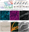
Fig. 3 |. Intercellular junctions in arachnoid and pia.
a. Region of meningeal layers in Fig. 2 showing junctions marked by lines at cell borders. All cells shown are interconnected by adherens junctions. Arachnoid barrier cells also have tight junctions and tricellular junctions that contribute to the permeability barrier of the layer, . b. Diagram illustrating the location and components of tricellular junctions between arachnoid barrier cells (from). Conventional bicellular tight junctions and adherens junctions are also present but are not shown for simplicity. c. Arachnoid barrier cells. Junctions in two overlapping layers are shown by staining of tight junction protein claudin-11, adherens junction protein cadherin-1/E-cadherin, and tricellular junction protein Lsr/angulin-1. Tricellular junctions (arrows) have strong staining for all three proteins. Some other junctions (arrowheads) have strong claudin-11 and E-cadherin but weak or no Lsr. Confocal microscopic images of adult mouse cerebral meningeal whole mount (from). d. Pia. Historical drawing showing the continuous layer of pial cells interconnected by junctions (black). Silver nitrate staining of dog pia (from). e. Pia. Intravital 2-photon image of mouse spinal cord showing VE-cadherin-GFP (green) at pial cell borders and blood vessels (orange) marked by fluorescent albumin (from). f. Pia. SEM image of stoma through which the subpial space is visible. Stoma (F). Cell nuclei (N). Macrophage (M). Dog cranial pia (from).

References

    1. Adeeb N, et al. The intracranial arachnoid mater : a comprehensive review of its history, anatomy, imaging, and pathology. Childs Nerv Syst 29, 17–33 (2013). - PubMed
    1. Engelhardt B, Vajkoczy P & Weller RO The movers and shapers in immune privilege of the CNS. Nature Immunology 18, 123–131 (2017). - PubMed
    1. Castro Dias M, Mapunda JA, Vladymyrov M & Engelhardt B Structure and Junctional Complexes of Endothelial, Epithelial and Glial Brain Barriers. Int J Mol Sci 20 (2019). - PMC - PubMed
    1. Derk J, Jones HE, Como C, Pawlikowski B & Siegenthaler JA Living on the Edge of the CNS: Meninges Cell Diversity in Health and Disease. Front Cell Neurosci 15, 703944 (2021). - PMC - PubMed
    1. Proulx ST Cerebrospinal fluid outflow: a review of the historical and contemporary evidence for arachnoid villi, perineural routes, and dural lymphatics. Cell Mol Life Sci 78, 2429–2457 (2021). - PMC - PubMed