. 2024 Sep 27;30(1):160.
doi: 10.1186/s10020-024-00924-6.

Pentagalloylglucose alleviates acetaminophen-induced acute liver injury by modulating inflammation via cGAS-STING pathway

Affiliations

Pentagalloylglucose alleviates acetaminophen-induced acute liver injury by modulating inflammation via cGAS-STING pathway

Congyang Zheng et al. Mol Med. .

Abstract

Background: The cGAS-STING pathway is an important component of the innate immune system and plays significant role in acetaminophen-induced liver injury (AILI). Pentagalloylglucose (PGG) is a natural polyphenolic compound with various beneficial effects, including anti-cancer, antioxidant, anti-inflammatory, and liver-protective properties; however, whether it can be used for the treatment of AILI and the specific mechanism remain unclear.

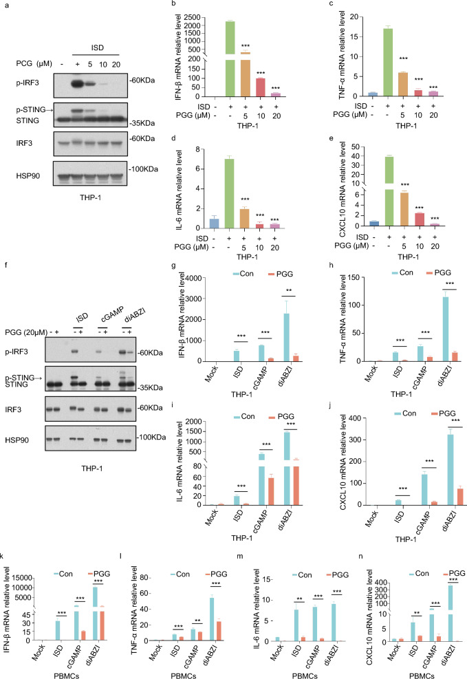
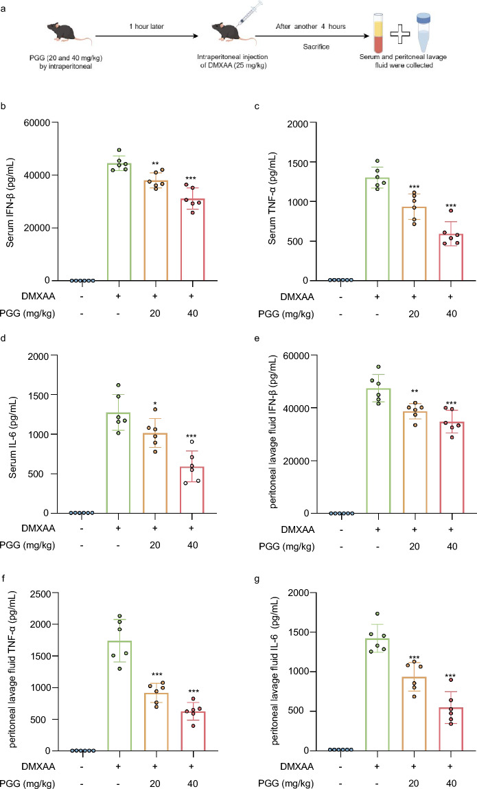
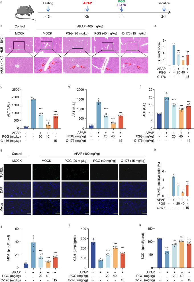
Materials and methods: A cell culture model was created to study the effect of PGG on cGAS-STING pathway activation using various techniques including western blotting (WB), real-time quantitative polymerase chain reaction (RT-qPCR), immunofluorescence (IF), and immunoprecipitation (IP). The effect of PGG was investigated in vivo by establishing a dimethylxanthenone acetic acid (DMXAA)-mediated activation model. An AILI model was used to evaluate the hepatoprotective and therapeutic effects of PGG by detecting liver function indicators, liver histopathology, and cGAS-STING pathway-related indicators in mice with AILI.

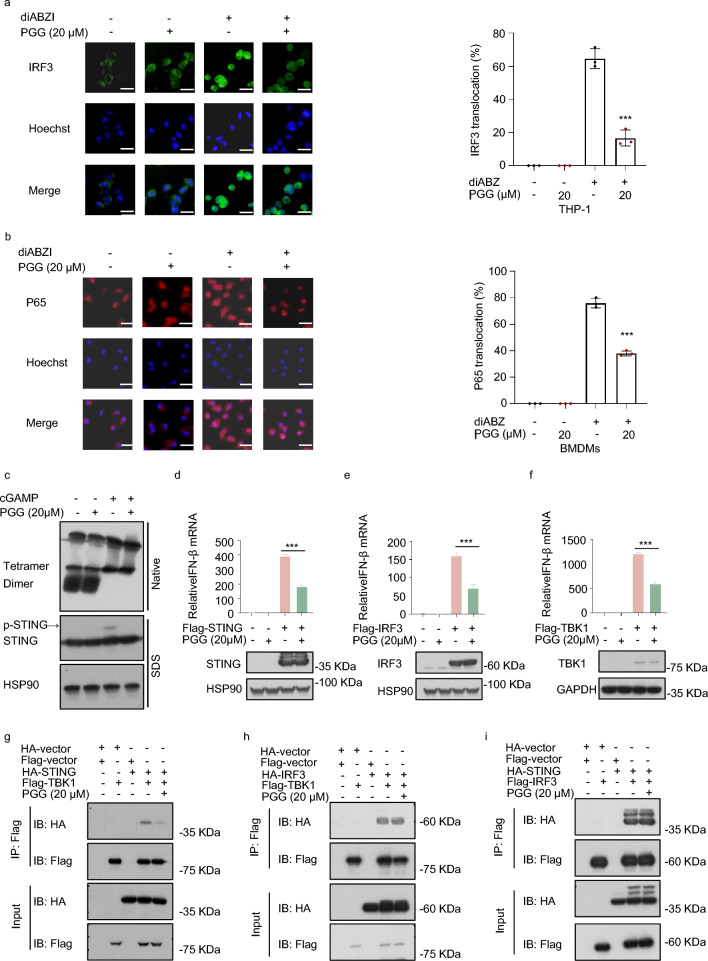
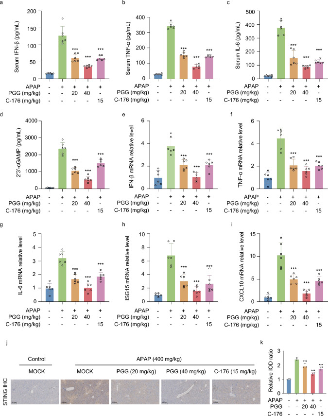
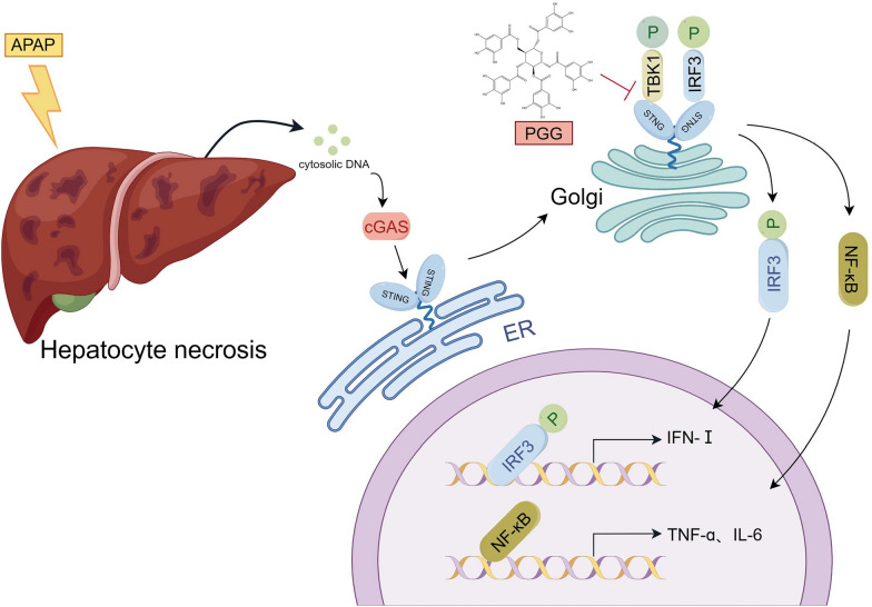
Results: PGG blocked cGAS-STING pathway activation in bone marrow-derived macrophages (BMDMs), THP-1 cells, and peripheral blood mononuclear cells (PBMCs) in vitro. Furthermore, PGG inhibited the generation of type I interferons (IFN-I) and the secretion of inflammatory factors in DMXAA-induced in vivo experiments. In addition, PGG also reduced serum levels of alanine aminotransferase (ALT), aspartate aminotransferase (AST), and alkaline phosphatase (ALP), improved liver tissue damage and apoptosis, and inhibited the cGAS-STING pathway activation caused by acetaminophen. In terms of the mechanism, PGG disrupted the connection between STING and TBK1.

Conclusions: PGG exerts a protective effect against AILI by blocking the cGAS-STING pathway, offering a promising treatment strategy.

Keywords: Acetaminophen; Acute liver injury; Pentagalloylglucose; cGAS-STING pathway.


Conflict of interest statement

The authors have stated that they do not have any conflicts of interest with any organization or individual that could potentially impact the outcomes and tendency of this research.

Figures

Fig. 1
Fig. 1
PGG inhibits cGAS-STING pathway activation in BMDMs. a BMDMs were pre-exposed with or without PGG (5, 10, 20 µM) 1 h before being activated by ISD for 2 h. Immunoblot analysis was performed to assess the levels of p-IRF3, IRF3, and STING. bf BMDMs were pre-exposed to either DMSO or PGG (5, 10, 20 µM) for 1 h before being activated with ISD for 4 h. The RT-qPCR analysis revealed the levels of IFN-β (b), TNF-α (c), IL-6 (d), ISG15 (e) and CXCL10 (f). g BMDMs were pretreated with or without PGG (5, 10, 20 µM) for 1 h and then stimulated with ISD, 2′3′-cGAMP, diABZI, or DMXAA for 2 h. Cell lysates were collected to detect the expression of relevant proteins. hl RT-qPCR assays for IFN-β (h), TNF-α (i), IL-6 (j), ISG15 (k) and CXCL10 (l) in BMDMs. The data were presented as means ± standard deviation (SD). **P < 0.01, ***P < 0.001. ns not significant
Fig. 2
Fig. 2
In THP-1 and PBMCs, PGG inhibits the activation of the cGAS-STING pathway. a PMA-primed THP-1 were pre-exposed with or without PGG (5, 10, 20 µM) for 1 h before being activated by ISD for 2 h. Immunoblotting was used to detect p-IRF3, IRF3, and STING. be PMA-primed THP-1 were pre-exposed with DMSO or PGG (5, 10, 20 µM) for 1 h before being activated with ISD for 4 h. The mRNA levels of IFN-β (b), TNF-α (c), IL-6 (d), and CXCL10 (e) were detected by RT-qPCR. f Western blot assays of p-IRF3, IRF3, and STING in PMA-primed THP-1. gj RT-qPCR assays for IFN-β (g), TNF-α (h), IL-6 (i), and CXCL10 (j) in PMA-primed THP-1. kn RT-qPCR was utilized to measure the expression levels of the IFN-β (k), TNF-α (l), IL-6 (m), and CXCL10 (n) genes in PBMCs. The data were presented as means ± SD. **P < 0.01, ***P < 0.001
Fig. 3
Fig. 3
PGG suppresses cGAS-STING pathway activation via inhibiting the interaction between STING and TBK1. a, b PMA-primed THP-1 or BMDMs were treated with DMSO or PGG at a concentration of 20 µM for 1 h, followed by stimulation with diABZI for 2 h. Immunostaining of IRF3 (a) and P65 (b) (n = 3). Scale bar, 10 μm. c After PGG at a concentration of 20 µM pretreatment for 1 h, BMDMs were stimulated with 2′3′-cGAMP for half an hour, followed by analysis of STING oligomerization and phosphorylation using immunoblotting. df Flag-tagged plasmid (Flag-STING, Flag-IRF3, Flag-TBK1) were transfected into HEK-293 cells for 12 h. Afterwards, cells were exposed to 20 μM PGG for a duration of 6 h and then subjected to WB and RT-qPCR. gi Flag-tagged plasmid (Flag-vector, Flag-TBK1, Flag-IRF3) and ha-tagged plasmid (HA-vector, HA-STING, HA-IRF3) were transfected into HEK-293 T cells for 24 h. Afterwards, cells were exposed to 20 μM PGG for a duration of 6 h and then subjected to immunoprecipitation using Anti-FLAG®M2Aftix gel for a period of 4 h in preparation for immunoblotting with the specified antibodies. The data were presented as means ± SD. ***P < 0.001
Fig. 4
Fig. 4
PGG inhibits the activation of STING and inflammatory response in vivo. a Schematic representation of an experiment performed on mice with DMXAA to induce STING activation. bd Mouse serum was collected to measure the levels of IFN-β (b), TNF-α (c) and IL-6 (d) using ELISA kits (n = 6). eg Mouse peritoneal lavage fluid was collected to measure the levels of IFN-β (e), TNF-α (f) and IL-6 (g) by ELISA kits (n = 6). The data were presented as means ± SD. *P < 0.05, **P < 0.01, ***P < 0.001 vs. the model group
Fig. 5
Fig. 5
PGG protect acetaminophen-induced acute liver injury. a Schematic illustration representing an experiment conducted on mice to induce acute liver injury using acetaminophen. b Representative H&E staining of liver tissues from AILI mice (n = 3). Scale bar: 100 µm. The red arrow points to the area of the liver lesion. (c) Grading the histological severity of liver injury induced by acetaminophen using the Suzuki score (n = 3). df Serum levels of ALT, AST and ALP from AILI mice (n = 6). g, h Representative images and quantification of TUNEL assay of liver tissues from AILI mice (n = 3). Scale bar: 100 µm. ik Hepatic tissue MDA, GSH and SOD levels were detected by biochemical kits (n = 6). The data were presented as means ± SD. ***P < 0.001 vs. the model group
Fig. 6
Fig. 6
PGG suppresses cGAS-STING Pathway activation in AILI mice. ac The levels of IFN-β, TNF-α and IL-6 from AILI mice serum were detected by ELISA kits (n = 6). d The levels of 2′3′-cGAMP from AILI mice liver tissues were detected by ELISA kits (n = 6). ei The mRNA levels of cGAS-STING pathway-related genes (IFN-β, TNF-α, IL-6, ISG15 and CXCL10) were measured by RT-qPCR assays in liver tissues from AILI mice (n = 6). jk Representative images and quantification of STING expression in liver tissues (n = 3). Scale bar: 100 µm. The data were presented as means ± SD. ***P < 0.001 vs. the model group
Fig. 7
Fig. 7
Graphic abstract. A schematic diagram showing that PGG attenuates AILI via inhibiting cGAS-STING pathway, involving suppressing binding between STING and TBK1

References

    1. Ashibe S, Ikeuchi K, Kume Y, Wakamori S, Ueno Y, Iwashita T, Yamada H. Non-enzymatic oxidation of a pentagalloylglucose analogue into members of the ellagitannin family. Angew Chem Int Ed Engl. 2017;56:15402–6. - PubMed
    1. Bakhoum SF, Ngo B, Laughney AM, Cavallo JA, Murphy CJ, Ly P, Shah P, Sriram RK, Watkins TBK, Taunk NK, et al. Chromosomal instability drives metastasis through a cytosolic DNA response. Nature. 2018;553:467–72. - PMC - PubMed
    1. Chen C, Xu P. Cellular functions of cGAS-STING signaling. Trends Cell Biol. 2023;33:630–48. - PubMed
    1. Chen L, Dong J, Liao S, Wang S, Wu Z, Zuo M, Liu B, Yan C, Chen Y, He H, et al. Loss of Sam50 in hepatocytes induces cardiolipin-dependent mitochondrial membrane remodeling to trigger mtDNA release and liver injury. Hepatology. 2022;76:1389–408. - PubMed
    1. Decout A, Katz JD, Venkatraman S, Ablasser A. The cGAS-STING pathway as a therapeutic target in inflammatory diseases. Nat Rev Immunol. 2021;21:548–69. - PMC - PubMed

MeSH terms