. 2024 Sep 27;15(1):329.
doi: 10.1186/s13287-024-03916-9.

Cytokine priming enhances the antifibrotic effects of human adipose derived mesenchymal stromal cells conditioned medium

Affiliations

Cytokine priming enhances the antifibrotic effects of human adipose derived mesenchymal stromal cells conditioned medium

Marianela Brizio et al. Stem Cell Res Ther. .

Abstract

Background: Fibrosis is a pathological scarring process characterized by persistent myofibroblast activation with excessive accumulation of extracellular matrix (ECM). Fibrotic disorders represent an increasing burden of disease-associated morbidity and mortality worldwide for which there are limited therapeutic options. Reversing fibrosis requires the elimination of myofibroblasts, remodeling of the ECM, and regeneration of functional tissue. Multipotent mesenchymal stromal cells (MSC) have antifibrotic properties mediated by secreted factors present in their conditioned medium (MSC-CM). However, there are no standardized in vitro assays to predict the antifibrotic effects of human MSC. As a result, we lack evidence on the effect of cytokine priming on MSC's antifibrotic effects. We hypothesize that the MSC-CM promotes fibrosis resolution in vitro and that this effect is enhanced following MSC cytokine priming.

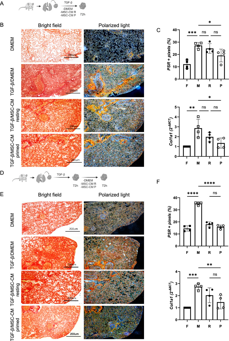
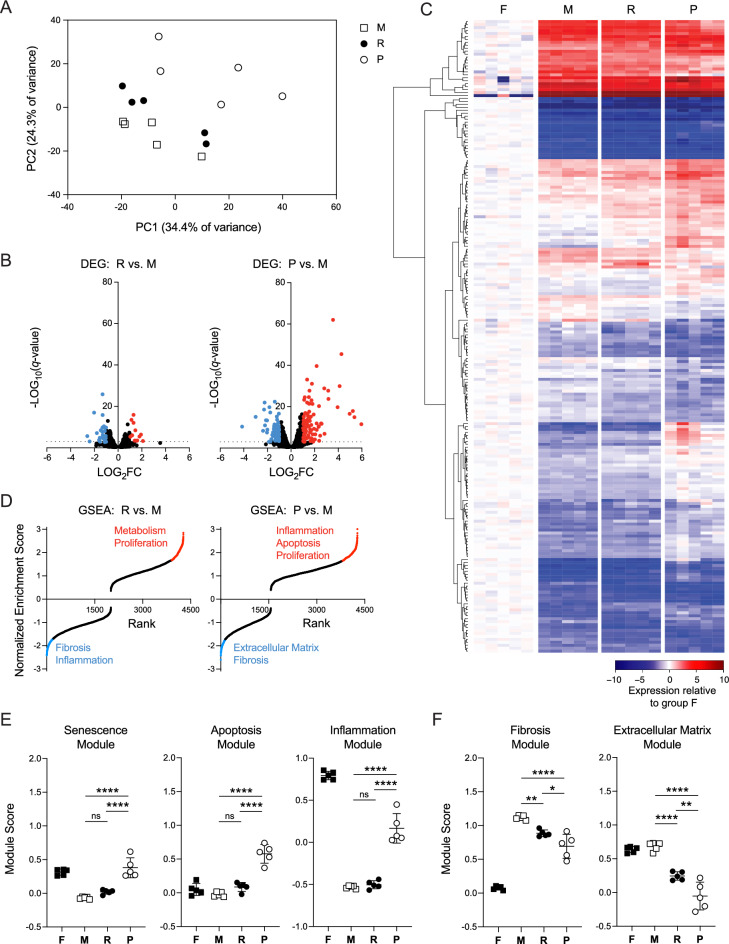
Methods: We compared the antifibrotic effects of resting versus interferon gamma (IFN-γ) and tumor necrosis factor alpha (TNF-α) primed MSC-CM in four in vitro assays: prevention of fibroblast activation, myofibroblasts deactivation, ECM degradation and fibrosis resolution in lung explant cultures. Furthermore, we performed transcriptomic analysis of myofibroblasts treated or not with resting or primed MSC-CM and proteomic characterization of resting and primed MSC-CM.

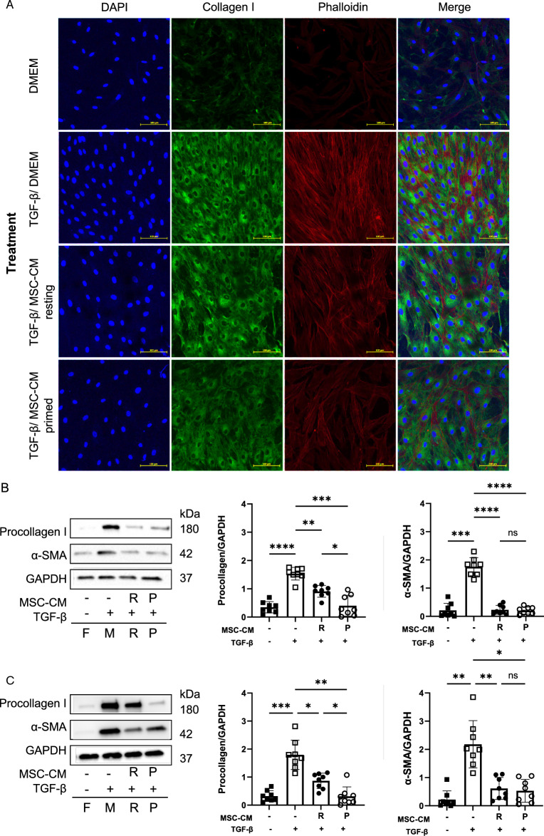
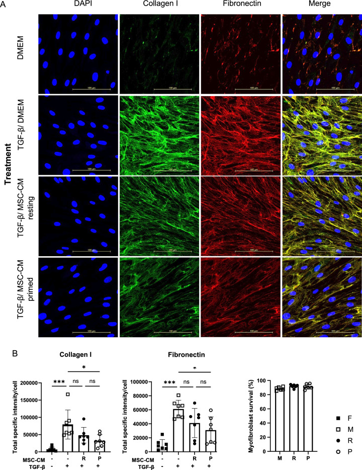
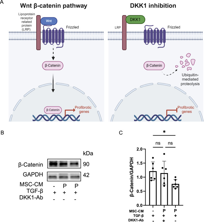
Results: We isolated MSC from adipose tissue of 8 donors, generated MSC-CM and tested each MSC-CM independently. We report that MSC-CM treatment prevented TGF-β induced fibroblast activation to a similar extent as nintedanib but, in contrast to nintedanib, MSC-CM reduced fibrogenic myofibroblasts (i.e. transcriptomic upregulation of apoptosis, senescence, and inflammatory pathways). These effects were larger when primed rather than resting MSC-CM were used. Priming increased the ability of MSC-CM to remodel the ECM, reducing its content of collagen I and fibronectin, and reduced the fibrotic load in TGF-β treated lung explant cultures. Priming increased the following antifibrotic proteins in MSC-CM: DKK1, MMP-1, MMP-3, follistatin and cathepsin S. Inhibition of DKK1 reduced the antifibrotic effects of MSC-CM.

Conclusions: In vitro, MSC-CM promote fibrosis resolution, an effect enhanced following MSC cytokine priming. Specifically, MSC-CM reduces fibrogenic myofibroblasts through apoptosis, senescence, and by enhancing ECM degradation. Future studies will establish the in vivo relevance of MSC priming to fibrosis resolution.

Keywords: Adipose tissue; Conditioned medium; Cytokine priming; Extracellular matrix; Fibroblasts; Fibrosis; Multipotent mesenchymal stromal cells; Myofibroblasts.


Conflict of interest statement

The authors declare that they have no competing interests.

Figures

Fig. 1
Fig. 1
Cytokine primed MSC-CM reduce fibroblast activation and promote myofibroblast deactivation in vitro. Fibroblasts were simultaneously treated with TGF-β and either resting (R) or cytokine primed (P) MSC-CM (i.e. prevention of TGF-β induced fibroblast activation assay). A Representative images showing reduction in intracellular collagen I (green) and stress fibers- phalloidin (red) in myofibroblasts treated with primed MSC-CM (scale: 100µm) and B Quantification of procollagen I and α-SMA by Western blot. C After 72 h of TGF-β activation, myofibroblasts were treated with either resting or primed MSC-CM (i.e. myofibroblast deactivation assay). Graphs depict the results of 8 independent experiments with means ± SD, ns = non-significant differences, *p < 0.05, **p < 0.01, ***p < 0.001. F: fibroblasts, M: myofibroblasts, R: myofibroblasts treated with resting MSC-CM, P: myofibroblasts treated with primed MSC-CM
Fig. 2
Fig. 2
Primed MSC-CM treatment changes the structure of extracellular collagen I and fibronectin. A Representative images showing collagen I (green) and fibronectin (red) secreted by myofibroblasts (scale: 100µm). After 5 days-treatment with primed MSC-CM. B Graphs summarizing the results from 7 independent experiments each with different MSC-CM. Each dot represents the average of 5 images. C Myofibroblast survival (annexin Vneg/DRAQ7.neg) was evaluated by flow cytometry (n = 6). ns = non-significant differences, *p < 0.05, **p < 0.01, ***p < 0.001
Fig. 3
Fig. 3
Primed MSC-CM treatment reduces fibrotic content in ex vivo lung explants. A–C Prevention of TGF-β explant activation assay: A Schematic workflow of the assay. B Representative images of negative controls (DMEM), positive controls (TGF-β + DMEM), and explants treated with MSC-CM resting (TGF-β + MSC-CM R) or primed MSC-CM (TGF-β + MSC-CM P). C Picrosirius red quantification (PSR) and collagen I gene expression. D-F Myofibroblast deactivation explant assay: D Schematic workflow of the assay. E Representative images of experimental conditions. F PSR quantification and collagen I gene expression. Graphs depict the results of 4 independent experiments with means ± SD. ns = non-significant differences, *p < 0.05, **p < 0.01, ***p < 0.001. F: fibroblasts, M: myofibroblasts, R: myofibroblasts treated with resting MSC-CM, P: myofibroblasts treated with primed MSC-CM
Fig. 4
Fig. 4
Transcriptional landscape of human fibroblasts treated TGF-β with and without resting or primed MSC-CM. Bulk RNA-seq was performed on fibroblasts (F), TGF-β-treated fibroblasts (myofibroblasts, M), and on myofibroblasts treated with resting MSC-CM (R) or with IFN-γ/TNF-α-primed MSC-CM (P) (n = 5 per group). A Principal component analysis (PCA) in M, R and P samples across 11,163 expressed genes at > 5 counts per million reads (CPM) in at least 3 samples. B Volcano plot of differentially expressed genes (DEG) in R versus M groups (left; 42 significant DEGs), and P versus M groups (right; 97 significant DEG), with a threshold of ± twofold change and q < 0.05 (Benjamini–Hochberg adjusted). C Heatmap of 214 total DEG calculated between groups M, R and P representing LOG2CPM gene expression in each sample expressed relative to group F. Hierarchical clustering of DEG was performed using Pearson correlation and Manhattan distance. D Normalized enrichment scores of canonical signaling pathway gene expression signatures using Gene Set Enrichment Analysis (GSEA). Positively (red) and negatively (blue) enriched gene sets met a cut-off of q < 0.05 (Benjamini–Hochberg adjusted). E, F Gene module scores calculated across all groups for E senescence, apoptosis, and inflammation gene signatures, and F fibrosis and extracellular matrix gene signatures. Data represent mean ± SD. Statistical test: One-way ANOVA with Tukey’s multiple correction test, * p < 0.05, ** p < 0.01, **** p < 0.0001
Fig. 5
Fig. 5
Priming changes the content of antifibrotic proteins in the MSC-CM. LC/MS–MS analysis of resting and primed MSC-CM. A Principal component analysis (PCA), (n = 4) B Venn diagrams indicating the total amount of identified proteins in MSC-CM (black = resting MSC-CM, white = primed MSC-CM) and C Heatmap of 43 proteins with a threshold of ± twofold change and p < 0.05 in resting and primed MSC-CM (red = present-high, blue = absent). Underlined are the proteins with known antifibrotic effects. D Antifibrotic proteins differentially present in primed MSC-CM. Multiplex analysis of E metalloproteases (MMP): MMP-1 and MMP-3 and F. tissue inhibitors of metalloproteases (TIMP) in resting and primed MSC-CM (n = 7). ns = non-significant differences, *p < 0.05. CTSS: Cathepsin S, FST: Follistatin, DKK1: Dickkopf-1, PROCR: Endothelial protein C receptor
Fig. 6
Fig. 6
DKK1 in primed MSC-CM inhibits the WNT pathway reducing β-catenin transcription. A Schematic representation of the WNT pathway and the inhibitory effect of DDK1 on β-catenin intracellular levels. B Western blot representative images and C summary graph of β-catenin abundance normalized by GAPDH intensity, (n = 6) ns = non-significant differences, *p < 0.05

References

    1. Atabai K, Yang CD, Podolsky MJ. You say you want a resolution (of fibrosis). Am J Respir Cell Mol Biol. 2020;63(4):424–35. - PMC - PubMed
    1. Thannickal VJ, et al. Fibrosis: ultimate and proximate causes. J Clin Investig. 2014;124(11):4673–7. - PMC - PubMed
    1. Henderson NC, Rieder F, Wynn TA. Fibrosis: from mechanisms to medicines. Nature. 2020;587(7835):555–66. - PMC - PubMed
    1. Rozier P, et al. Extracellular vesicles are more potent than adipose mesenchymal stromal cells to exert an anti-fibrotic effect in an in vitro model of systemic sclerosis. Int J Mol Sci. 2021;22(13):6837. - PMC - PubMed
    1. Hall MC, et al. The comparative role of activator protein 1 and Smad factors in the regulation of Timp-1 and MMP-1 gene expression by transforming growth factor-beta 1. J Biol Chem. 2003;278(12):10304–13. - PubMed

MeSH terms