. 2024 Sep 27;17(1):69.
doi: 10.1186/s13041-024-01144-z.

Electroacupuncture inhibited carrageenan-induced pain aversion by activating GABAergic neurons in the ACC

Affiliations

Electroacupuncture inhibited carrageenan-induced pain aversion by activating GABAergic neurons in the ACC

Yichen Zhu et al. Mol Brain. .

Abstract

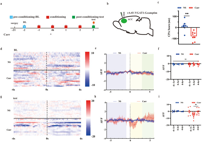
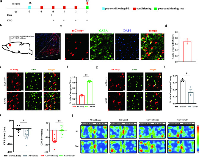
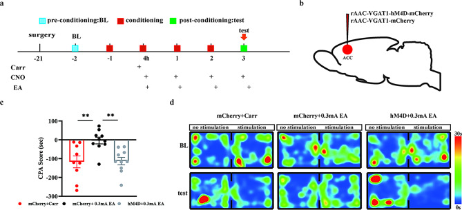
Pain aversion is an avoidance response to painful stimuli. Previous research has indicated that the anterior cingulate cortex (ACC) is involved in pain aversion processing. However, as interneurons, the role of GABAergic neurons in the ACC (GABAACC neurons) in pain aversion is still unclear. Electroacupuncture (EA) has been shown to ameliorate pain aversion, but the mechanism is not clarified. The present study provided evidence that inhibition of GABAACC neurons contributed to pain aversion. EA alleviated pain aversion by activating GABAACC neurons in an intensity-dependent manner. Specifically, 0.3 mA EA stimulation showed better effects on pain aversion than 0.1 mA stimulation, which could be reversed by chemical genetic inhibition of GABAACC neurons. These results provide a novel mechanism by which EA alleviates pain aversion by reversing GABAACC neurons.

Keywords: Anterior cingulate cortex; Electroacupuncture; GABAergic neurons; Pain; Pain aversion.


Conflict of interest statement

The authors declare that the study was conducted without any commercial or financial relationships that could be considered a potential conflict of interest.

Figures

Fig. 1
Fig. 1
Injection of carrageenan induced pain aversion (A) Flow chart for this part of the experiment. (B) Comparison of the left-paw withdrawal thresholds of the NS group and Carr group at different times. **P < 0.01 compared with the NS group. (C) CPA scores of mice in the NS group and Carr gro(D) up. Representative graphs of CPA thermograms for each group
Fig. 2
Fig. 2
The ACC is involved in the formation of pain aversion. (A) Flow chart of the experiment. (B) Mechanical pain threshold of the saline + NS, saline + Carr and QA + Carr groups. Compared with the saline + NS group, **P < 0.01; compared with the saline + Carr group, ##P < 0.01. (C) CPA scores of the saline + NS, saline + Carr and QA + Carr groups. Pairwise comparisons **P < 0.01. (D) Schematic diagram of drug destruction in ACC brain regions, scale bar, 500 μm
Fig. 3
Fig. 3
Decreased activity of GABAACC neurons in carrageenan-induced inflammatory pain model mice induced by mechanical pain stimulation. (A) Flow chart of the experiment. (B) Sagittal map and location diagram of virus injection and optical fiber implantation, scale bar, 100 μm. (C) Representative ΔF/F heat chart (from one mouse, 5 trials) of activity changes in ACC GABAergic neurons before and after Carr injection. (D) Representative ΔF/F line chart (from one mouse, 5 trials) of activity changes in ACC GABAergic neurons before and after Carr injection. (E) AUC diagram (0–10 s) of the activity of GABAACC neurons before and after Carr injection, pairwise comparisons, *P < 0.05. (n = 4 mice)
Fig. 4
Fig. 4
Changes in the activity of ACC GABAergic neurons during the formation of pain aversion. (A) Flow chart of the experiment. (B) Sagittal map of virus and optical fiber implantation. (C) CPA scores of mice in the experiment. Compared with the NS group, **P < 0.01. (D) Thermography of GABAergic neurons in the NS group and Carr group before and after entering the pain box at baseline. (E-F) ΔF/F line graph and area under the curve of ΔF/F at the baseline state. (G) Thermography of GABAergic neurons in the NS group and Carr group before and after entering the pain box under test conditions. (H-I) ΔF/F line graph and area under the curve of ΔF/F in the test state, pairwise comparisons, *P < 0.05
Fig. 5
Fig. 5
Effect of chemical genetic modulation of GABAACC neurons on pain aversion. (A) Flow chart of the experiment. (B) Schematic of chemog(C) enetic virus injection (left), schematic of mCherry-labeled cells (right) scale bar, 100 μm. Colocalization of mCherry (red) and GABA in the ACC brain region, scale bar, 20 μm. (D) Percentage of mCherry and GABA colocalization (n = 15, 3 mice) GABA green (E) and (G) ACC Representative maps of GABAergic neurons colabeled with c-Fos, c-Fos green, GABAergic neurons red. Scale bars, 20 μm. (F) and (H) Percentage of ACC GABAergic neurons colabeled with c-Fos (n = 15, 3 mice). Pairwise comparisons, **P < 0.01. (I) Experimental results of CPA in the NS + mCherry group, NS + hM4D group, Carr + mCherry group and Carr + hM3D group, pairwise comparisons, **P < 0.01. (J) Representative graphs of CPA th(J) ermograms for each group
Fig. 6
Fig. 6
The effect of different intensities of EA on pain aversion behaviors. (A) Flow chart of the experiment. (B) Results of conditioned CPA in each group, pairwise comparisons, **P < 0.01. (C) Representative heatmap of conditioned aversion experiments in the Carr group, 0.3 mA EA group, and 0.1 mA EA group. (D) Colocalization of GABA (green) with DAPI (blue) in the ACC brain region in each group (n = 15, 3 mice). Scale bar, 50 μm. (E) Statistical map of the number of GABA neurons in the ACC in each group, pairwise comparisons, **P < 0.01
Fig. 7
Fig. 7
EA regulates pain aversion behaviors through GABAACC neurons. (A) Flow chart of the experiment. (B) Schematic diagram of viral injection sites. (C) CPA results of mice in the mCherry + Carr group, mCherry + 0.3 mA EA group, and hM4D + 0.3 mA EA group; pairwise comparisons, **P < 0.01. (D) Representative heatmap of the conditioned aversion experiment for each group

Similar articles

References

    1. Treede RD, Rief W, Barke A, Aziz Q, Bennett MI, Benoliel R, et al. Chronic pain as a symptom or a disease: the IASP classification of Chronic Pain for the International classification of diseases (ICD-11). Pain. 2019;160:19–27. - PubMed
    1. Cohen SP, Vase L, Hooten WM. Chronic pain: an update on burden, best practices, and new advances. Lancet. 2021;397:2082–97. - PubMed
    1. Clauw DJ, Essex MN, Pitman V, Jones KD. Reframing chronic pain as a disease, not a symptom: rationale and implications for pain management. Postgrad Med. 2019;131:185–98. - PubMed
    1. Treede RD, Rief W, Barke A, Aziz Q, Bennett MI, Benoliel R, et al. A classification of chronic pain for ICD-11. Pain. 2015;156:1003–7. - PMC - PubMed
    1. Rogers AH, Orr MF, Shepherd JM, Bakhshaie J, Ditre JW, Buckner JD, et al. Anxiety, depression, and opioid misuse among adults with chronic pain: the role of emotion dysregulation. J Behav Med. 2021;44:66–73. - PMC - PubMed

LinkOut - more resources