. 2024 Sep 27;22(1):862.
doi: 10.1186/s12967-024-05650-5.

An oncolytic HSV-1 vector induces a therapeutic adaptive immune response against glioblastoma

Affiliations

An oncolytic HSV-1 vector induces a therapeutic adaptive immune response against glioblastoma

Alberto Reale et al. J Transl Med. .

Abstract

Background: Glioblastoma (GBM) is the most frequent and aggressive brain tumor in adults with the lowest survival rates five years post-diagnosis. Oncolytic viruses (OVs) selectively target and damage cancer cells, and for this reason they are being investigated as new therapeutic tools also against GBM.

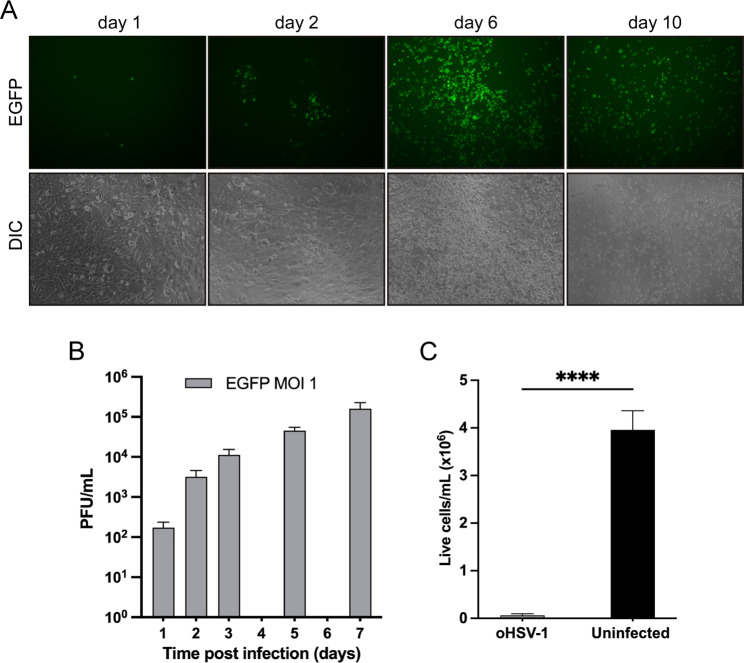
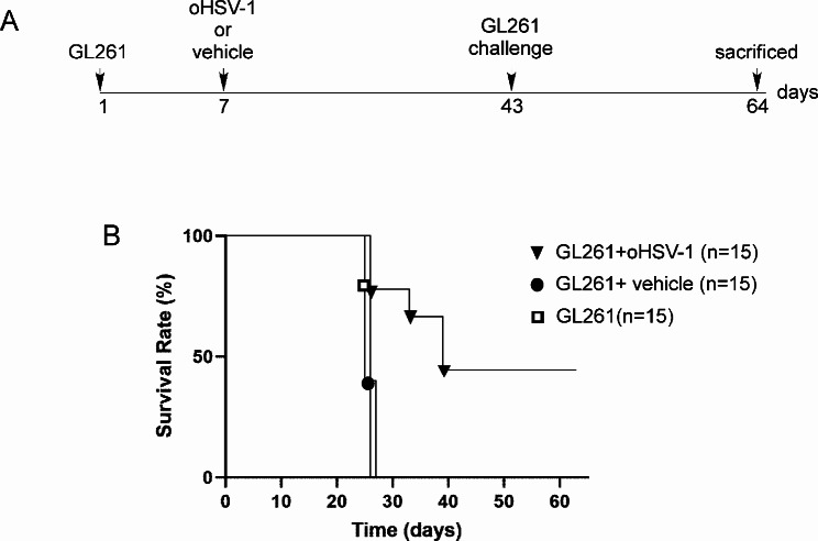
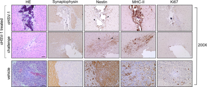
Methods: An oncolytic herpes simplex virus type 1 (oHSV-1) with deletions in the γ34.5 neurovirulence gene and the US12 gene, expressing enhanced green fluorescent protein (EGFP-oHSV-1) as reporter gene was generated and tested for its capacity to infect and kill the murine GL261 glioblastoma (GBM) cell line. Syngeneic mice were orthotopically injected with GL261cells. Seven days post-implantation, EGFP-oHSV-1 was administered intratumorally. Twenty-one days after parental tumor challenge in the opposite brain hemisphere, mice were sacrified and their brains were analysed by immunohistochemistry to assess tumor presence and cell infiltrate.

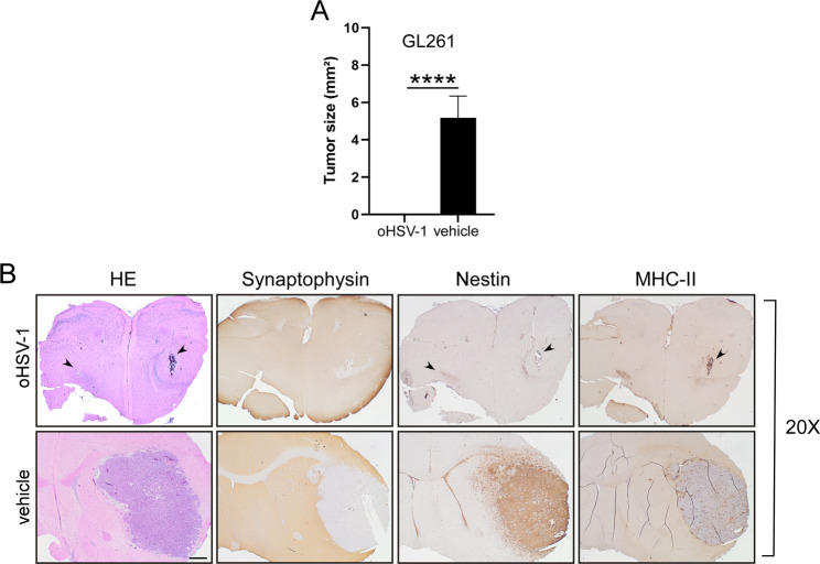
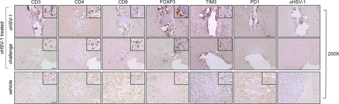
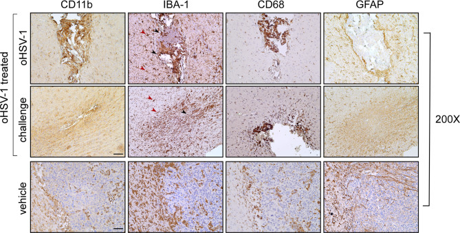
Results: oHSV-1 replicates and induces cell death of GL261 cells in vitro. A single intracranial injection of EGFP-oHSV-1 in established GL261 tumors significantly prolongs survival in all treated mice compared to placebo treatment. Notably, 45% of treated mice became long-term survivors, and rejected GL261 cells upon rechallenge in the contralateral brain hemisphere, indicating an anamnestic antitumoral immune response. Post-mortem analysis revealed a profound modification of the tumor microenvironment with increased infiltration of CD4 + and CD8 + T lymphocytes, intertumoral vascular collapse and activation and redistribution of macrophage, microglia, and astroglia in the tumor area, with the formation of intense fibrotic tissue suggestive of complete rejection in long-term survivor mice.

Conclusions: EGFP-oHSV1 demonstrates potent antitumoral activity in an immunocompetent GBM model as a monotherapy, resulting from direct cell killing combined with the stimulation of a protective adaptive immune response. These results open the way to possible application of our strategy in clinical setting.

Keywords: GL261; Glioblastoma; HSV-1; Immunotherapy; Oncolytic virus.


Conflict of interest statement

The authors declare that they have no competing interests.

Figures

Fig. 1
Fig. 1
oHSV-1 replicates and induces cell death in GL261 glioblastoma cells in vitro. (A) GL261 cells were seeded in a 24-well plate (7.5 × 104 cells/well), infected with oHSV-1 (MOI = 1) for 1 h in serum-free DMEM medium, then maintained in DMEM medium supplemented with 2% fetal bovine serum (FBS). The medium was refreshed daily, and EGFP expression, serving as an indicator of viral infection, was monitored at 1, 2, 6, and 10 days post-infection. Fluorescence was monitored using a Leica EpiFluorescence microscope revealing a progressive spread of the infection to the entire monolayer. Concurrently, brightfield microscopy demonstrated an increasing cytopathic effect, characterized by cellular rounding. All images were captured at 10x magnification and represent multiple microscopic fields from a triplicate experiment. (B) GL261 cells were seeded in a 24-well plate (7.5 × 104 cells/well), infected with oHSV-1 (MOI = 1) for 1 h in serum-free DMEM medium, then maintained in DMEM medium supplemented with 2% FBS. The supernatant was collected and replaced with fresh 2% FBS medium every 24 h. Infectious viral particles were quantified as plaque forming units (PFU)/mL by plaque titration assay on green monkey Vero cells. The experiment was performed in triplicate. Y axis is in logarithmic scale. (C) 10 days after infection, surviving infected GL261 cells were trypsinized and counted using a hemocytometer and Trypan blue exclusion staining. Uninfected GL261 cells seeded and cultured in the same conditions for 10 days were counted in a similar way. The experiment was performed in triplicate and the difference in the number of live cells between uninfected and oHSV-1-infected GL261 cells was evaluated by Student’s t-test. ****P < 0.0001. In all panels, error bars represent standard deviation
Fig. 2
Fig. 2
oHSV-1 treatment of GL261 tumor-bearing mice significantly improve survival. (A) Experimental setup and treatment schedule is depicted. (B) oHSV-1 intratumoral treatment (black triangles) significantly improved survival compared to animals treated with vehicle (black circles) or left untreated (white squares) with median survival of 38, 26 and 26 days respectively (P < 0.05). Survival was analyzed by Kaplan–Meier method and compared by log-rank Mantel-Cox test
Fig. 3
Fig. 3
Intracranial injection of oHSV-1 in established GL261 tumor dramatically blocks tumor growth in vivo and rejects GL261 tumor challenge in the opposite brain hemisphere. C57BL/6 mice (n = 45) were initially injected with 3 × 104 GL261 cells into the right striatum. After seven days of tumor implantation, one group of mice injected with GL261 cells was further treated with 106 plaque-forming units (PFU) of oHSV-1 in the same brain hemisphere (n = 15). As a control, another group of mice received intracranial injections of PBS (vehicle) (n = 15) and the other animals were left untreated (n = 15). At 43 post-tumor implantation all surviving mice (n = 7) were challenged with parental 3 × 104 GL261 tumor cells in the opposite brain hemisphere, and after additional 21 days mice were sacrificed, brains were removed, and serial sections of the brain were carried out to measure tumor size and for staining. (A) Average tumor size of GL261 tumor in oHSV-1 treated mice (oHSV-1) and in control group (vehicle). Data are represented as mean values, and error bars indicate the standard deviation (SD) within each group. p-Values were determined via unpaired t-test; ****P < 0.0001. (B) HE and IHC staining of serial brain sections. The panels were taken at 20x magnification. Arrowheads in HE, Nestin and MHC-II upper panels (oHSV-1) point to the sites of tumor cell injection shwing the absence of growing tumors. HE, hematoxylin and eosin; IHC, immunohistochemistry. Scale bar corresponds to 500 μM
Fig. 4
Fig. 4
Immunohistological characterization of GL261 tumor rejection in oHSV-1 treated mice. Representative histological sections of tumor tissues harvested from pre-treated (oHSV-1 treated: oHSV-1 and challenge) and un-treated mice (vehicle) mice, at 200x magnification. HE was followed by immunohistochemical staining with synaptophysin- and nestin-specific antibodies to better identify tumoral and non-tumoral tissue, respectively. Arrowheads in the panel showing nestin staining of oHSV-1 treated mice point to astrocytosis-enriched areas in both brain hemispheres. MHC-II expression was observed on myeloid cells concentrated over the tumor bed in both brain hemispheres of oHSV-1 treated mice (oHSV-1 treated) or dispersed along the GL261 tumor (vehicle). Ki67-positive cells are abundant in tumors isolated from non-treated mice (vehicle). HE, hematoxylin and eosin. Scale bar corresponds to 50 μM
Fig. 5
Fig. 5
Rejected GL261 parental tumors in oHSV-1 pre-treated mice are strongly infiltrated by both CD4 + and CD8 + T cells and do not present HSV-1 viral particles. Representative immunohistology images of tumor sections. Slides of brain tissues isolated from oHSV-1-injected GL261 tumors (oHSV-1) and challenged with GL261 (challenge) are indicated in the upper and middle panels, respectively. Slides of brain tissues isolated from GL261 + vehicle (vehicle) are indicated in the lower panels. Sections were stained by IHC with anti-CD3, anti-CD4, anti-CD8, anti-FOXP3, anti-TIM3, anti-PD1, and anti-HSV-1 antibodies. Small square boxes are the areas represented in the corresponding large square boxes of each IHC image. Images were taken at 200x magnification. Large square boxes were taken at 400x magnification. IHC, immunohistochemistry. Scale bar corresponds to 50 μM
Fig. 6
Fig. 6
Immunohistological characterization of tumor microenvironment in GL261 C57BL/6 bearing mice treated with oHSV-1 compared to untreated mice. IHC staining of serial brain sections. Slides of brain tissues isolated from oHSV-1-injected GL261 tumors and challenged with GL261 are indicated in the upper and middle panels, respectively. Slides of brain tissues isolated from GL261-untreated mice are represented in the lower panels (vehicle). Sections were stained by IHC with anti-CD11b, anti-CD68, anti-IBA1 and anti-GFAP antibodies. Images were taken at 200x magnification. Black and red aarrowheads in the IBA-1-stained upper and middle panels point to ameboid and branched macrophages, respectively. IBA-1: Ionized calcium-binding adapter molecule 1, GFAP: glial fibrillary acidic protein. Scale bar corresponds to 50 μM

References

    1. Keller BA, Bell JC. Oncolytic viruses-immunotherapeutics on the rise. J Mol Med (Berl). 2016;94:979–91. - PubMed
    1. Campadelli-Fiume G, Petrovic B, Leoni V, Gianni T, Avitabile E, Casiraghi C, et al. Retargeting strategies for oncolytic herpes simplex viruses. Viruses. 2016;8:63. - PMC - PubMed
    1. Kaufman HL, Kohlhapp FJ, Zloza A. Oncolytic viruses: a new class of immunotherapy drugs. Nat Rev Drug Discov. 2015;14:642–62. - PMC - PubMed
    1. Conry RM, Westbrook B, McKee S, Norwood TG. Talimogene laherparepvec: first in class oncolytic virotherapy. Hum Vaccin Immunother. 2018;14:839–46. - PMC - PubMed
    1. Lasner TM, Tal-Singer R, Kesari S, Lee VM, Trojanowski JQ, Fraser NW. Toxicity and neuronal infection of a HSV-1 ICP34.5 mutant in nude mice. J Neurovirol. 1998;4:100–5. - PubMed

Substances

LinkOut - more resources