. 2024 Sep 27;19(1):130.
doi: 10.1186/s13000-024-01552-8.

Aberrant positivity for BCOR immunohistochemistry in merkel cell carcinoma - a potential diagnostic pitfall

Affiliations

Aberrant positivity for BCOR immunohistochemistry in merkel cell carcinoma - a potential diagnostic pitfall

Viola Katharina Vetter et al. Diagn Pathol. .

Abstract

Backrgound: Merkel cell carcinoma (MCC) is a rare, aggressive primary cutaneous neuroendocrine carcinoma, frequently associated with clonal Merkel cell polyomavirus integration. MCC can pose significant diagnostic challenges due to its diverse clinical presentation and its broad histological differential diagnosis. Histologically, MCC presents as a small-round-blue cell neoplasm, where the differential diagnosis includes basal cell carcinoma, melanoma, hematologic malignancies, round cell sarcoma and metastatic small cell carcinoma of any site. We here report strong aberrant immunoreactivity for BCOR in MCC, a marker commonly used to identify round cell sarcomas and other neoplasms with BCOR alterations.

Methods: Based on strong BCOR expression in three index cases of MCC, clinically mistaken as sarcoma, a retrospective analysis of three patient cohorts, comprising 31 MCC, 19 small cell lung carcinoma (SCLC) and 5 cases of neoplasms with molecularly confirmed BCOR alteration was conducted. Immunohistochemical staining intensity and localization for BCOR was semi-quantitatively analyzed.

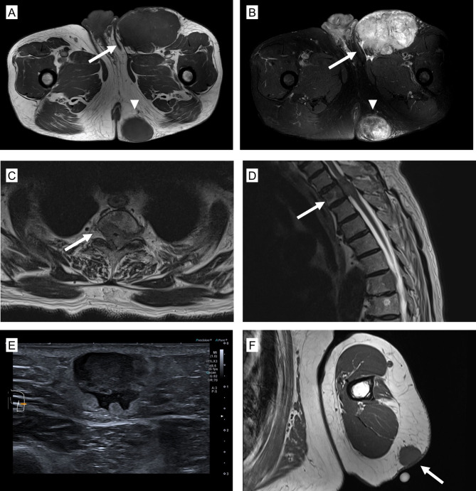
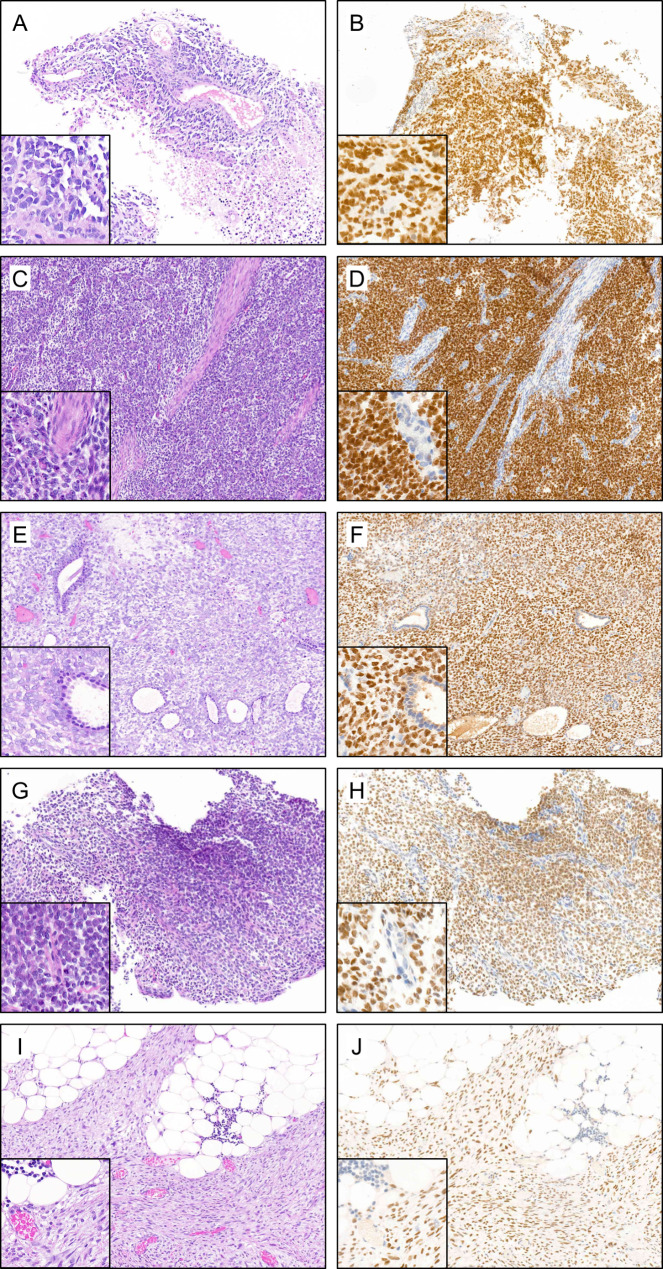
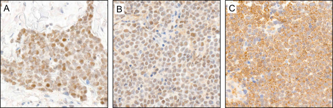
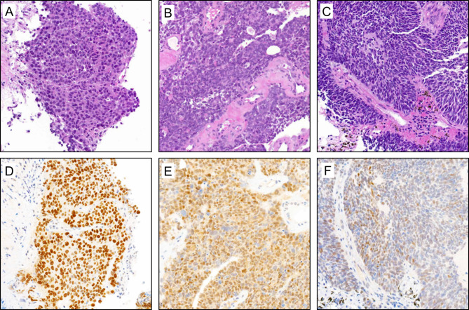
Results: Three cases, clinically and radiologically mimicking a sarcoma were analyzed in our soft tissue and bone pathology service. Histologically, the cases showed sheets of a small round blue cell neoplasm. A broad panel of immunohistochemistry was used for lineage classification. Positivity for synaptophysin, CK20 and Merkel cell polyoma virus large T-antigen lead to the diagnosis of a MCC. Interestingly, all cases showed strong positive nuclear staining for BCOR, which was included for the initial work-up with the clinical differential of a round cell sarcoma. We analyzed a larger retrospective MCC cohort and found aberrant weak to strong BCOR positivity (nuclear and/or cytoplasmic) in up to 90% of the cases. As a positive control, we compared the expression to a small group of BCOR-altered neoplasms. Furthermore, we investigated a cohort of SCLC as another neuroendocrine neoplasm and found in all cases a diffuse moderate to strong BCOR positivity.

Conclusions: This study demonstrates that neuroendocrine neoplasms, such as MCC and SCLC can express strong aberrant BCOR. This might represent a diagnostic challenge or pitfall, in particular when MCC is clinically mistaken as a soft tissue or a bone sarcoma.

Keywords: BCOR; Immunohistochemistry; Merkel cell carcinoma; Neuroendocrine carcinoma; SCLC; Sarcoma.


Conflict of interest statement

All authors have no conflict of interest.

Figures

Fig. 1
Fig. 1
Radiologic findings of three MCC cases presenting as a soft tissue / bone mass (A), axial T1-weighted, and (B), axial T2-weighted fat-saturated MRI images of case 1 showing two lesions in the inguinal region (arrow) and gluteal region (arrowhead) with T1-isointensity to muscle (A) and T2-hyperintensity (B). C, axial, and D, sagittal, T2-weighted MRI images of case 2 showing a slightly hypointense lesion in the second thoracic vertebra (arrow) in the transversal (C) and sagittal plane (D) and additional similar lesions in other thoracic vertebrae. (E, F) Diagnostic imaging of case 3 showing a lesion on the upper arm. (E) Ultrasound image depicting a hypoechogenic, partially ill-defined mass. (F) axial T1-weighted MRI image showing a mass (arrow) with isointensity to muscle
Fig. 2
Fig. 2
Histology and immunohistochemistry of three MCC cases presenting as a soft tissue / bone mass (A-C) HE stains showing sheets of a small-round-blue cell neoplasm. (D-F): CK20 immunohistochemistry (brown) showing diffuse or focal dot-like (F) staining. (G-I): Positivity for Synaptophysin (brown). (J-L): Positivity for MCPyV (red). (M-O): strong nuclear positivity for BCOR (brown)
Fig. 3
Fig. 3
HE and BCOR staining in tumors with molecularly confirmed BCOR-alterations: All cases show diffuse nuclear moderate to strong BCOR staining. Inlets show higher magnification. (A, B and C, D): sarcomas with BCOR-ITD. (E, F): sarcoma with a BCOR::CNNB3 fusion. (G, H): clear cell sarcoma of the kidney with BCOR-ITD. (I, J): high-grade endometrial stroma sarcoma with an EP300::BCOR fusion
Fig. 4
Fig. 4
BCOR staining patterns in MCC: (A) moderate nuclear staining (B) moderate nuclear staining accompanied by a weak cytoplasmic staining (C) peri-nuclear dot-like staining
Fig. 5
Fig. 5
HE and BCOR staining in 3 SCLC cases: (A-C): HE staining of three SCLC cases. (D-F): Corresponding BCOR immunohistochemistry. The first case (A, D) shows strong, diffuse nuclear staining. The second case (B, E) shows moderate to strong nuclear and weak cytoplasmic staining. The third case (C, F) shows partial moderate nuclear staining

References

    1. Heath M, Jaimes N, Lemos B, Mostaghimi A, Wang LC, Penas PF, Nghiem P. Clinical characteristics of Merkel cell carcinoma at diagnosis in 195 patients: the AEIOU features. J Am Acad Dermatol. 2008;58(3):375–81. - PMC - PubMed
    1. van der Zwan JM, Trama A, Otter R, Larranaga N, Tavilla A, Marcos-Gragera R, et al. Rare neuroendocrine tumours: results of the surveillance of rare cancers in Europe project. Eur J Cancer. 2013;49(11):2565–78. - PubMed
    1. Youlden DR, Soyer HP, Youl PH, Fritschi L, Baade PD. Incidence and survival for Merkel cell carcinoma in Queensland, Australia, 1993–2010. JAMA Dermatol. 2014;150(8):864–72. - PubMed
    1. Harms KL, Healy MA, Nghiem P, Sober AJ, Johnson TM, Bichakjian CK, Wong SL. Analysis of prognostic factors from 9387 Merkel cell carcinoma cases forms the basis for the New 8th Edition AJCC Staging System. Ann Surg Oncol. 2016;23(11):3564–71. - PMC - PubMed
    1. Astolfi A, Fiore M, Melchionda F, Indio V, Bertuccio SN, Pession A. BCOR involvement in cancer. Epigenomics. 2019;11(7):835–55. - PMC - PubMed

MeSH terms