. 2024 Sep 27;15(1):325.
doi: 10.1186/s13287-024-03946-3.

Human uncultured adipose-derived stromal vascular fraction shows therapeutic potential against osteoarthritis in immunodeficient rats via direct effects of transplanted M2 macrophages

Affiliations

Human uncultured adipose-derived stromal vascular fraction shows therapeutic potential against osteoarthritis in immunodeficient rats via direct effects of transplanted M2 macrophages

Yuma Onoi et al. Stem Cell Res Ther. .

Abstract

Background: The uncultured adipose-derived stromal vascular fraction (SVF), consisting of adipose-derived stromal cells (ADSCs), M2 macrophages (M2Φ) and others, has shown therapeutic potential against osteoarthritis (OA), however, the mechanisms underlying its therapeutic effects remain unclear. Therefore, this study investigated the effects of the SVF on OA in a human-immunodeficient rat xenotransplantation model.

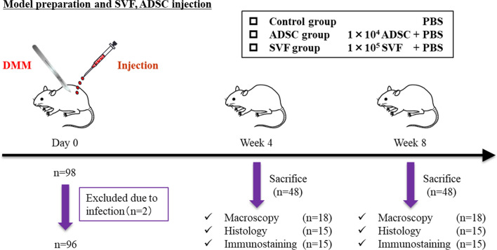
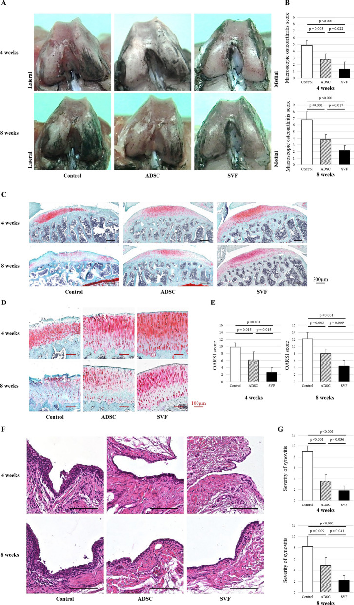
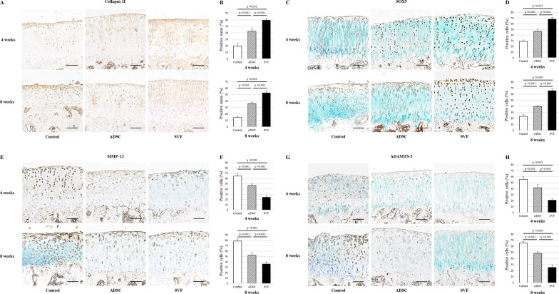
Methods: OA model was induced in the knees of female immunodeficient rats by destabilization of the medial meniscus. Immediately after the surgery, human SVF (1 × 10⁵), ADSCs (1 × 10⁴), or phosphate buffered saline as a control group were transplanted into the knees. At 4 and 8 weeks postoperatively, OA progression and synovitis were analyzed by macroscopic and histological analyses, and the expression of collagen II, SOX9, MMP-13, ADAMTS-5, F4/80, CD86 (M1), CD163 (M2), and human nuclear antigen (hNA) were evaluated immunohistochemically. In vitro, flow cytometry was performed to collect CD163-positive cells as M2Φ from the SVF. Chondrocyte pellets (1 × 10⁵) were co-cultured with SVF (1 × 10⁵), M2Φ (1 × 10⁴), and ADSCs (1 × 10⁴) or alone as a control group, and the pellet size was compared. TGF-β, IL-10 and MMP-13 concentrations in the medium were evaluated using enzyme-linked immunosorbent assay.

Results: In comparison with the control and ADSC groups, the SVF group showed significantly slower OA progression and less synovitis with higher expression of collagen II and SOX9, lower expression of MMP-13 and ADAMTS-5, and lower F4/80 and M1/M2 ratio in the synovium. Only the SVF group showed partial expression of hNA-, CD163-, and F4/80-positive cells in the rat synovium. In vitro, the SVF, M2Φ, ADSC and control groups, in that order, showed larger pellet sizes, higher TGF-β and IL-10, and lower MMP-13 concentrations.

Conclusions: The M2Φ in the transplanted SVF directly affected recipient tissue, enhancing the secretion of growth factors and chondrocyte-protecting cytokines, and partially improving chondrocytes and joint homeostasis. These findings indicate that the SVF is as an effective option for regenerative therapy for OA, with mechanisms different from those of ADSCs.

Keywords: Adipose-derived stromal cells; M2 macrophages; Osteoarthritis; Rat model; Stromal vascular fraction; Synovitis; Xenotransplantation.


Conflict of interest statement

The authors have no competing interests to declare.

Figures

Fig. 1
Fig. 1
A schematic diagram illustrating the study design, the time course of the study, and the number of rats at each stage. SVF, stromal vascular fraction; ADSC, adipose-derived stromal cell; PBS, phosphate-buffered saline; DMM, destabilization of the medial meniscus
Fig. 2
Fig. 2
A Macroscopic analysis of femoral condyles showing typical specimens at 4 and 8 weeks after DMM. The cartilage surface was stained with India ink to confirm fibrosis and erosion. B Macroscopic osteoarthritis scores at 4 and 8 weeks after DMM in each group (n = 6/group). C, D Histological analysis of tibial cartilage. Safranin-O/fast green staining of typical specimens at 4 and 8 weeks after DMM in each group. Scale bar = 300 μm or 100 μm. E OARSI scores at 4 and 8 weeks after DMM in each group (n = 5/group). F Histological analysis of synovium. Hematoxylin and eosin staining of typical specimens at 4 and 8 weeks after DMM in each group. Scale bar = 100 μm. G Modified OARSI synovitis scores at 4 and 8 weeks after DMM in each group (n = 5/group). SVF, stromal vascular fraction; ADSC, adipose-derived stromal cell; DMM, destabilization of the medial meniscus; OARSI, Osteoarthritis Research Society International
Fig. 3
Fig. 3
A Immunohistochemical analysis of collagen II in cartilage showing representative samples at 4 and 8 weeks after DMM. Scale bar = 100 μm. B Percentage of collagen II-positive areas at 4 and 8 weeks after DMM in each group (n = 5/group). C Immunohistochemical analysis of SOX9 in cartilage showing representative samples at 4 and 8 weeks after DMM. Scale bar = 100 μm. D Percentage of SOX9-positive cells at 4 and 8 weeks after DMM in each group (n = 5/group). E Immunohistochemical analysis of MMP-13 in cartilage showing representative samples at 4 and 8 weeks after DMM. Scale bar = 100 μm. F Percentage of MMP-13-positive cells at 4 and 8 weeks after DMM in each group (n = 5/group). G Immunohistochemical analysis of ADAMTS-5 in cartilage showing representative samples at 4 and 8 weeks after DMM. Scale bar = 100 μm. H Percentage of ADAMTS-5-positive cells at 4 and 8 weeks after DMM in each group (n = 5/group). SVF, stromal vascular fraction; ADSC, adipose-derived stromal cell; DMM, destabilization of the medial meniscus; SOX9, SRY box 9; MMP-13, matrix metalloproteinases 13; ADAMTS-5, A disintegrin and metalloproteinase with thrombospondin motifs 5
Fig. 4
Fig. 4
A Immunohistochemical analysis of F4/80 in synovial tissue showing representative samples at 4 and 8 weeks after DMM. The sections were counterstained with hematoxylin. Scale bar = 100 μm. B F4/80 scores in each group (n = 5/group). C Immunohistochemical analysis of CD86 in synovial tissue showing representative samples at 4 and 8 weeks after DMM. The sections were counterstained with hematoxylin. Scale bar = 100 μm. D Immunohistochemical analysis of CD163 in synovial tissue showing representative samples at 4 and 8 weeks after DMM. The sections were counterstained with hematoxylin. Scale bar = 100 μm. (E) CD86/CD163 expression ratio (M1/M2 ratio) in each group (n = 5/group). SVF, stromal vascular fraction; ADSC, adipose-derived stromal cell; DMM, destabilization of the medial meniscus; MΦ, macrophages
Fig. 5
Fig. 5
A Immunofluorescence analysis of F4/80, CD163, and hNA in synovial tissue of the rats at 4 weeks after DMM showing representative triple immunostaining of F4/80 (green), CD163 (red), and hNA (violet) in each group. Scale bar = 20 μm. B Comparison of the ratio of CD163- and hNA-positive cells in the F4/80-positive cells via immunofluorescence staining (n = 5/group). SVF, stromal vascular fraction; ADSC, adipose-derived stromal cell; hNA, human nuclear antigen
Fig. 6
Fig. 6
A Flow cytometry analysis revealing the proportion of CD163-positive cells in the SVF (n = 5). B Setup of the separated pellet co-culture system. Groups including ADSC, M2Φ, and SVF were established, with each administered cell type in membrane plates and OA chondrocytes in 15 mL tubes. A control group was established with no cells in membrane plates and only OA chondrocytes in 15-mL tubes. C Gross photographs and safranin-O staining of the resulting pellets in each group. D Comparison of pellet size among each group (n = 5/group). E Analysis of TGF-β, IL-10, and MMP-13 levels in the supernatant following coculture with ADSC, M2Φ, and SVF and the chondrocyte (n = 5/group). SVF, stromal vascular fraction; M2Φ, M2 macrophages; ADSC, adipose-derived stromal cell; TGF-β, transforming growth factor-β; IL-10, interleukin-10; MMP-13, matrix metalloproteinase 13

References

    1. Agarwal N, Mak C, Bojanic C, To K, Khan W. Meta-analysis of adipose tissue derived cell-based therapy for the treatment of knee osteoarthritis. Cells. 2021;10(6):1365. - PMC - PubMed
    1. Zuk PA, Zhu M, Mizuno H, Huang J, Futrell JW, Katz AJ, et al. Multilineage cells from human adipose tissue: implications for cell-based therapies. Tissue Eng. 2001;7(2):211–28. - PubMed
    1. Feng Z, Ting J, Alfonso Z, Strem BM, Fraser JK, Rutenberg J, et al. Fresh and cryopreserved, uncultured adipose tissue-derived stem and regenerative cells ameliorate ischemia–reperfusion-induced acute kidney injury. Nephrol Dial Transplant. 2010;25(12):3874–84. - PMC - PubMed
    1. Li M, Lei H, Xu Y, Li H, Yang B, Yu C, et al. Exosomes derived from mesenchymal stem cells exert therapeutic effect in a rat model of cavernous nerves injury. Andrology. 2018;6(6):927–35. - PubMed
    1. Pomatto M, Gai C, Negro F, Cedrino M, Grange C, Ceccotti E, et al. Differential therapeutic effect of extracellular vesicles derived by bone marrow and adipose mesenchymal stem cells on wound healing of diabetic ulcers and correlation to their cargoes. Int J Mol Sci. 2021;22(8):3851. - PMC - PubMed