. 2024 Sep 27;55(1):124.
doi: 10.1186/s13567-024-01373-z.

Development and characterization of reverse genetics systems of feline infectious peritonitis virus for antiviral research

Affiliations

Development and characterization of reverse genetics systems of feline infectious peritonitis virus for antiviral research

Guoqian Gu et al. Vet Res. .

Erratum in

Abstract

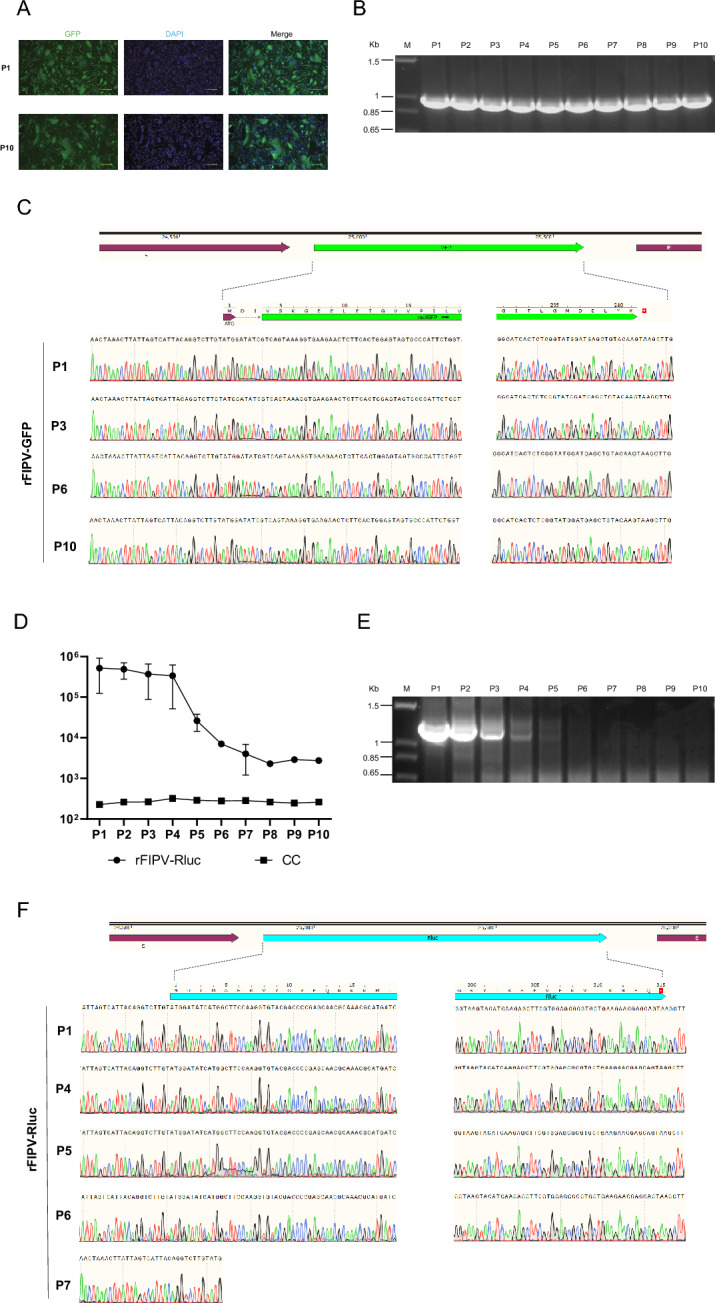
Feline infectious peritonitis (FIP) is a lethal, immune-mediated disease in cats caused by feline infectious peritonitis virus (FIPV), a biotype of feline coronavirus (FCoV). In contrast to feline enteric coronavirus (FECV), which exclusively infects enterocytes and causes diarrhea, FIPV specifically targets macrophages, resulting in the development of FIP. The transmission and infection mechanisms of this complex, invariably fatal disease remain unclear, with no effective vaccines or approved drugs for its prevention or control. In this study, a full-length infectious cDNA clone of the wild-type FIPV WSU79-1149 strain was constructed to generate recombinant FIPV (rFIPV-WT), which exhibited similar growth kinetics and produced infectious virus titres comparable to those of the parental wild-type virus. In addition, the superfold green fluorescent protein (msfGFP) and Renilla luciferase (Rluc) reporter genes were incorporated into the rFIPV-WT cDNA construct to generate reporter rFIPV-msfGFP and rFIPV-Rluc viruses. While the growth characteristics of the rFIPV-msfGFP virus were similar to those of its parental rFIPV-WT, the rFIPV-Rluc virus replicated more slowly, resulting in the formation of smaller plaques than did the rFIPV-WT and rFIPV-msfGFP viruses. In addition, by replacing the S, E, M, and ORF3abc genes with msfGFP and Rluc genes, the replicon systems repFIPV-msfGFP and repFIPV-Rluc were generated on the basis of the cDNA construct of rFIPV-WT. Last, the use of reporter recombinant viruses and replicons in antiviral screening assays demonstrated their high sensitivity for quantifying the antiviral effectiveness of the tested compounds. This integrated system promises to significantly streamline the investigation of virus replication within host cells, enabling efficient screening for anti-FIPV compounds and evaluating emerging drug-resistant mutations within the FIPV genome.

Keywords: Feline infectious peritonitis virus; antiviral compounds; high-content screening; recombinant viruses; replicon; reverse genetics.


Conflict of interest statement

The authors declare that they have no competing interests.

Figures

Figure 1
Figure 1
Construction and characterization of the rFIPV-msfGFP and rFIPV-Rluc viruses. A Schematic diagram showing the genomic structures of the rFIPV-WT, rFIPV-msfGFP, and rFIPV-Rluc viruses used in this study and the strategy for constructing the recombinant viruses. The ORF3abc genes in the FIPV genome were replaced with msfGFP and Rluc genes. The full-length FIPV genome was divided into seven fragments. The seven fragments and the T7 promoter were subsequently cloned and inserted into the PCR-XL-2 vector by the TOPO cloning assay, as a BsaI restriction site flanked each fragment. The PCR-XL-2 carrying each fragment was digested with BsaI and ligated with the T4 ligation enzyme. The full-length genomic cDNA and N gene cDNA were transcribed into RNA in vitro and transfected into CRFK cells via electroporation. The schematic diagram is not drawn to scale. B Morphology and size of viral plaques of the rFIPV-WT, rFIPV-msfGFP, and rFIPV-Rluc strains compared with those of the parental FIPV WSU79-1146 strain. Representative images of the plaque morphology of the above three recombinant viruses and the wild-type FIPV WSU79-1146 are shown. The areas of 30 plaques were determined and plotted for each virus. The experiment was repeated three times with similar results, and the results of one representative experiment are shown. ***p < 0.001. C Growth kinetics of the recombinant viruses. CRFK cells were infected with recombinant viruses and wild-type FIPV WSU79-1146 at an MOI of 0.1. Viral titres from culture supernatants at the indicated time points were determined by plaque assays. The error bars indicate the means and standard deviations from three independent experiments.
Figure 2
Figure 2
Genetic stability of the rFIPV-msfGFP and rFIPV-Rluc viruses. The rFIPV-msfGFP and rFIPV-Rluc viruses were serially passaged at an MOI of 0.1 in CRFK cells ten times. A Representative fluorescence images of rFIPV-msfGFP virus-infected CRFK cells during passage. P1 and P10 are shown. B Analysis of the genetic stability of the rFIPV-msfGFP virus after ten passages. Viral RNA was extracted from the culture supernatants of each passage, and RT-PCR was performed with a primer set flanking the msfGFP gene. The resulting RT-PCR products were resolved by 1% agarose gel electrophoresis. The 1-kb DNA ladders are indicated. C Chromatograms of Sanger sequencing results of the msfGFP gene of the rFIPV-msfGFP virus from P1, 3, 6, and 10 are shown. D Rluc activity of rFIPV-Rluc at each passage. For each passage, the cell lysate samples were subjected to luminescence analysis. Uninfected cells served as the cell control (CC). The error bars indicate the means and standard deviations from three independent experiments. E Analysis of the genetic stability of the rFIPV-Rluc virus after ten passages. Viral RNA was extracted from culture supernatants of each passage, and RT-PCR was performed with a primer set flanking the Rluc gene. The resulting RT-PCR products were resolved by 1% agarose gel electrophoresis. The 1-kb DNA ladders are indicated. F Chromatograms of Sanger sequencing results of the Rluc gene of the rFIPV-Rluc virus from P1, 4, 5, 6, and 7 are shown.
Figure 3
Figure 3
Application of the rFIPV-msfGFP and rFIPV-Rluc viruses in drug screening. A Antiviral assay of GS-441524 or GC-376 using wild-type FIPV and their cytotoxic effects. Antiviral assay of GS-441524 or GC-376 using B rFIPV-msfGFP and C rFIPV-Rluc. Relative inhibition was calculated on the basis of reporter gene expression in cells treated with antiviral drugs compared with that in cells treated with DMSO. EC50 values and CC50 values were calculated as explained in the Materials and Methods and are represented on individual graphs. The error bars indicate the means and standard deviations from three independent experiments.
Figure 4
Figure 4
Construction of repFIPV-msfGFP and repFIPV-Rluc. A Schematic diagram showing the genomic structures of the FIPV replicons used in this study and the strategy for constructing the replicons. The FIPV replicon genomic cDNA and N gene cDNA were transcribed into RNA in vitro and transfected into CRFK cells via electroporation. B Kinetics of msfGFP gene expression postelectroporation in CRFK cells. The number of GFP-positive cells in each well containing 4 × 104 cells was counted at the indicated time points. The error bars indicate the means and standard deviations from three independent experiments. C Kinetics of Rluc gene expression post-electroporation in CRFK cells. For each time point, the cell lysate samples were subjected to luminescence analysis. The untransfected cells served as the CC. The error bars indicate the means and standard deviations from three independent experiments.
Figure 5
Figure 5
Application of FIPV replicons in drug discovery. Dose-dependent responses of A repFIPV-msfGFP and B repFIPV-Rluc reporter activity to GS-441524, GC-376, and E64d. Inhibition was determined by the percentage of reporter gene signals compared with that in DMSO-treated cells. The cytotoxicity of GS-441524, GC-376, and E64d to the cells was also examined. EC50 values and CC50 values were calculated as explained in the Materials and Methods and are represented on individual graphs. The error bars indicate the means and standard deviations from three independent experiments.

Similar articles

Cited by

References

    1. Woo PCY, de Groot RJ, Haagmans B, Lau SKP, Neuman BW, Perlman S, Sola I, van der Hoek L, Wong ACP, Yeh SH (2023) ICTV virus taxonomy profile: coronaviridae 2023. J Gen Virol 104:001843 - PubMed
    1. Tekes G, Thiel HJ (2016) Feline coronaviruses: pathogenesis of feline infectious peritonitis. Adv Virus Res 96:193–218 - PMC - PubMed
    1. Li C, Liu Q, Kong F, Guo D, Zhai J, Su M, Sun D (2019) Circulation and genetic diversity of Feline coronavirus type I and II from clinically healthy and FIP-suspected cats in China. Transbound Emerg Dis 66:763–775 - PMC - PubMed
    1. Benetka V, Kubber-Heiss A, Kolodziejek J, Nowotny N, Hofmann-Parisot M, Mostl K (2004) Prevalence of feline coronavirus types I and II in cats with histopathologically verified feline infectious peritonitis. Vet Microbiol 99:31–42 - PMC - PubMed
    1. Amer A, Siti Suri A, Abdul Rahman O, Mohd HB, Faruku B, Saeed S, Tengku Azmi TI (2012) Isolation and molecular characterization of type I and type II feline coronavirus in Malaysia. Virol J 9:278 - PMC - PubMed

LinkOut - more resources