. 2024 Sep 2;13(9):1072.
doi: 10.3390/antiox13091072.

Beneficial Effect of Dimethyl Fumarate Drug Repositioning in a Mouse Model of TDP-43-Dependent Frontotemporal Dementia

Affiliations

Beneficial Effect of Dimethyl Fumarate Drug Repositioning in a Mouse Model of TDP-43-Dependent Frontotemporal Dementia

Ignacio Silva-Llanes et al. Antioxidants (Basel). .

Abstract

Frontotemporal dementia (FTD) causes progressive neurodegeneration in the frontal and temporal lobes, leading to behavioral, cognitive, and language impairments. With no effective treatment available, exploring new therapeutic approaches is critical. Recent research highlights the transcription factor Nuclear Factor erythroid-derived 2-like 2 (NRF2) as vital in limiting neurodegeneration, with its activation shown to mitigate FTD-related processes like inflammation. Dimethyl fumarate (DMF), an NRF2 activator, has demonstrated neuroprotective effects in a TAU-dependent FTD mouse model, reducing neurodegeneration and inflammation. This suggests DMF repositioning potential for FTD treatment. Until now, no trial had been conducted to analyze the effect of DMF on TDP-43-dependent FTD. In this study, we aimed to determine the potential therapeutic efficacy of DMF in a TDP-43-related FTD mouse model that exhibits early cognitive impairment. Mice received oral DMF treatment every other day from presymptomatic to symptomatic stages. By post-natal day (PND) 60, an improvement in cognitive function is already evident, becoming even more pronounced by PND90. This cognitive enhancement correlates with the neuroprotection observed in the dentate gyrus and a reduction in astrogliosis in the stratum lacunosum-moleculare zone. At the prefrontal cortex (PFC) level, a neuroprotective effect of DMF is also observed, accompanied by a reduction in astrogliosis. Collectively, our results suggest a potential therapeutic application of DMF for patients with TDP-43-dependent FTD.

Keywords: DMF; NRF2; TDP-43; neurodegeneration; neuroinflammation.


Conflict of interest statement

The authors declare no conflicts of interest.

Figures
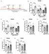
Figure 1
Figure 1
Response in the Novel Object Recognition (NOR) test of CAMKII-TDP-43 and WT mice at PND60 and PND90 after a chronic i.g. administration of DMF (100 mg/kg) from PND45 up to PND90 (A) Timeline representation of the experimental design: At PND45, we started the treatments (VEH, or DMF 100 mg/kg, i.g., respectively). At PND60, we performed the first NOR analysis (four consecutive days), and at PND90, before sacrifice, we performed another NOR test. (B,C) Analysis of the exploration time of familiar object (TF) and novel object (TN) at PND60 and PND90. (D,E) Analysis of the discrimination index at PND60 and PND90. (F,G) Analysis of the recognition index at PND60 and PND90. Bars indicate the mean of n = 10–12 samples ± SEM. Asterisks show significant differences with * p < 0.05, ** p < 0.01, *** p < 0.005, **** p < 0.001 comparing each group according to a one-way ANOVA followed by Tukey’s post-test.
Figure 2
Figure 2
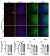
DMF has a neuroprotective effect on the granular cell layer of the dentate gyrus. (A) Immunofluorescence staining of DAPI and CALBINDIN-D28K of 30 μm-thick sections of the dentate gyrus of the hippocampus from WT, and CAMKII-TDP-43 mice treated with vehicle or DMF. (B) Quantification of the area stained with DAPI in the dentate gyrus from WT, and CAMKII-TDP-43 mice treated with vehicle or DMF. (C) Quantification of the intensity of the area stained with CALBINDIN-D28K in the dentate gyrus from WT, and CAMKII-TDP-43 mice treated with vehicle or DMF. (D) Quantification of the area stained with CALBINDIN-D28K in the dentate gyrus from WT, and CAMKII-TDP-43 mice treated with vehicle or DMF. Bars indicate the mean of n = 4–5 samples ± SEM. Asterisks show significant differences with * p < 0.05 comparing each group according to a one-way ANOVA followed by Tukey’s post-test.
Figure 3
Figure 3
DMF treatment modulates the astrogliosis observed in CAMKII-TDP-43 mice at the hippocampus. (A) Immunofluorescence of IBA1 (red) and GFAP (green), microglial and astrocytic markers, respectively, of 30 μm-thick sections in the CA1-hippocampus of mice treated with VEH or DMF, n = 4–5 samples ± SEM. Quantification of number of microglial (B) and astrocytes (C) cells at the CA1 area of mice treated with VEH or DMF, n = 4–5 samples ± SEM. RT-qPCR determination of mRNA levels of Trem2 (D), Il1b (E), Glast1 (F), and Sphk2 (G) genes at the hippocampus of mice treated with VEH or DMF, n = 4–5 samples ± SEM. The asterisks represent the difference in significance: * p < 0.05, ** p < 0.01, comparing each group according to a one-way ANOVA followed by Tukey’s post-test.
Figure 4
Figure 4
TDP-43 overexpression decreases CTIP2+ neurons and DMF treatment partially reverses this effect in layer V of the cortex. (A) Immunofluorescence of CTIP2 (marker of corticospinal motor neurons and other projection neurons in layer V) (red) and DAPI (blue) of 30 μm-thick sections of the mPFC of mice treated with VEH or DMF, n = 5 samples ± SEM. (B) Quantification of number of CTIP2+ cells at the layer V of the mPFC of mice treated with VEH or DMF, n= 4–5 samples ± SEM. The asterisks represent the difference in significance: ** p < 0.01, comparing each group according to a one-way ANOVA followed by Tukey’s post-test.
Figure 5
Figure 5
The overexpression of TDP-43 induces astrogliosis in layer V of the mPFC and treatment with DMF partially reverses this effect. (A) Immunofluorescence of IBA1 (red) and S100B (green), microglial and astrocytic markers, respectively, of 30 μm-thick sections in the layer V of the mPFC of mice treated with VEH or DMF, n = 4–5 samples ± SEM. Quantification of number of microglial (B) and astrocyte (C) cells at the layer V of the mPFC of mice treated with VEH or DMF, n = 4–5 samples ± SEM. RT-qPCR determination of mRNA levels of Il1b (D) and Sphk2 (E) genes in the same area, n = 4–5 samples ± SEM. The asterisks represent the difference in significance: * p < 0.05, ** p < 0.01, comparing each group according to a one-way ANOVA followed by Tukey’s post-test.
Figure 6
Figure 6
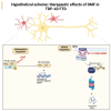
Hypothetical scheme of the neuroprotective effect of DMF treatment in a TDP-43-dependent FTD model. Our results suggest an NRF2-dependent effect mediated by the actions of DMF and MMF at the antioxidant level and an anti-inflammatory effect, which may be primarily mediated by DMF.

References

    1. Leroy M., Bertoux M., Skrobala E., Mode E., Adnet-Bonte C., Le Ber I., Bombois S., Cassagnaud P., Chen Y., Deramecourt V., et al. Characteristics and progression of patients with frontotemporal dementia in a regional memory clinic network. Alzheimer’s Res. Ther. 2021;13:19. doi: 10.1186/s13195-020-00753-9. - DOI - PMC - PubMed
    1. Grossman M., Seeley W.W., Boxer A.L., Hillis A.E., Knopman D.S., Ljubenov P.A., Miller B., Piguet O., Rademakers R., Whitwell J.L., et al. Frontotemporal lobar degeneration. Nat. Rev. Dis. Primers. 2023;9:40. doi: 10.1038/s41572-023-00447-0. - DOI - PubMed
    1. Tong K.I., Padmanabhan B., Kobayashi A., Shang C., Hirotsu Y., Yokoyama S., Yamamoto M. Different electrostatic potentials define ETGE and DLG motifs as hinge and latch in oxidative stress response. Mol. Cell. Biol. 2007;27:7511–7521. doi: 10.1128/MCB.00753-07. - DOI - PMC - PubMed
    1. Itoh K., Chiba T., Takahashi S., Ishii T., Igarashi K., Katoh Y., Oyake T., Hayashi N., Satoh K., Hatayama I., et al. An Nrf2/small Maf heterodimer mediates the induction of phase II detoxifying enzyme genes through antioxidant response elements. Biochem. Biophys. Res. Commun. 1997;236:313–322. doi: 10.1006/bbrc.1997.6943. - DOI - PubMed
    1. Itoh K., Wakabayashi N., Katoh Y., Ishii T., Igarashi K., Engel J.D., Yamamoto M. Keap1 represses nuclear activation of antioxidant responsive elements by Nrf2 through binding to the amino-terminal Neh2 domain. Genes. Dev. 1999;13:76–86. doi: 10.1101/gad.13.1.76. - DOI - PMC - PubMed

LinkOut - more resources