. 2024 Sep 4;14(9):1122.
doi: 10.3390/biom14091122.

The Role of Claudins in the Pathogenesis of Dextran Sulfate Sodium-Induced Experimental Colitis: The Effects of Nobiletin

Affiliations

The Role of Claudins in the Pathogenesis of Dextran Sulfate Sodium-Induced Experimental Colitis: The Effects of Nobiletin

Asmaa Al-Failakawi et al. Biomolecules. .

Abstract

Background: The pathogenesis of inflammatory bowel diseases such as ulcerative colitis and Crohn's disease is not well understood. This study investigated the roles and regulation of the claudin-1, -2, -3, and -4 isoforms in the pathogenesis of ulcerative colitis, and the potential therapeutic effects of nobiletin.

Methods: Colitis was induced in rats by administering dextran sulfate sodium [DSS] in drinking water for seven days. Animals were treated daily with nobiletin [oral, 60 mg/Kg body weight] and studied in four groups, C [non-colitis control], D [DSS-induced colitis], CN [nobiletin-treated non-colitis control], and DN [nobiletin-treated DSS-induced colitis]. On day seven, the animals were sacrificed, and colonic tissues were collected and analyzed.

Results: Both macroscopic and microscopic findings suggest the progression of colitis. In the inflamed colon, claudin-1 and -4 proteins were decreased, claudin-2 increased, while the claudin-3 protein remained unchanged. Except for claudin-1, these changes were not paralleled by mRNA expression, indicating a complex regulatory mechanism. Uniform β-actin expression along with consistent quality and yield of total RNA indicated selectivity of these changes. Nobiletin treatment reversed these changes.

Conclusions: Altered expression of the claudin isoforms -1, -2, and -4 disrupts tight junctions, exposing the lamina propria to microflora, leading to electrolyte disturbance and the development of ulcerative colitis. Nobiletin with its anti-inflammatory properties may be useful in IBD.

Keywords: claudins; colitis; dextran sulfate sodium; inflammation; myeloperoxidase; tight junctions.


Conflict of interest statement

The authors declare that they have no conflicts of interest.

Figures
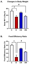
Figure 1
Figure 1
(A) Body weight (Wt) change [percentage of day 0 body weight] and (B) the food efficiency ratios of the animals from the test conditions: C [non-colitis control], D [DSS-induced colitis], CN [nobiletin-treated non-colitis control], and DN [nobiletin-treated DSS-induced colitis]. The data are the mean ± SE [n = 10]. *, # indicate significance at p < 0.05 between the indicated pair of groups.
Figure 2
Figure 2
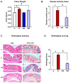
(A) Colon weights [g/cm colon length], (B) the disease activity score, (C) a representative histology of hematoxylin and eosin-stained [left panel] and alcian blue-stained [right panel] colonic tissue sections, and (D) the histological score of the animals from the test conditions: C [non-colitis control], D [DSS-induced colitis], CN [nobiletin-treated non-colitis control], and DN [nobiletin-treated DSS-induced colitis]. The data are the mean ± SE [n = 10]. *, # indicate significance at p < 0.05 between the indicated pair of groups. Magnification: 10×. Scalebar = 100 μm.
Figure 3
Figure 3
A bar diagram showing MPO activity units/min-mg in the colonic segments from the test conditions: C [non-colitis control], D [DSS-induced colitis], CN [nobiletin-treated non-colitis control], and DN [nobiletin-treated DSS-induced colitis]. The data are the mean ± SE [n = 10]. *, # indicate significance at p < 0.05 between the indicated groups.
Figure 4
Figure 4
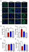
(A) Representative photomicrographs showing immunofluorescence staining, and (BE) a bar diagram showing the expression levels of the indicated claudin isoforms measured as mean fluorescence intensity [MFI, arbitrary units] using a specific primary antibody and an FITC-labeled secondary antibody in the tissue sections of colon from the test conditions: C [non-colitis control], D [DSS-induced colitis], CN [nobiletin-treated non-colitis control], and DN [nobiletin-treated DSS-induced colitis]. The tissue sections were counter-stained with DAPI. Claudins are expressed in the surface lining epithelium [arrows] and in the epithelial cells [arrow heads] lining the glands [G]. L—lumen of the colon. Magnification = 20×, Scalebars = 100 μm. The data are the mean ± SE [n = 10]. *, # indicate significance at p < 0.05 between the indicated pair of groups.
Figure 5
Figure 5
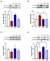
(A) A bar diagram showing the protein expression levels relative to β-actin [ratios] of claudin-1, (B) claudin-2, (C) claudin-3, and (D) claudin-4 isoforms in the colon from the test conditions: C [non-colitis control], D [DSS-induced colitis], CN [nobiletin-treated non-colitis control], and DN [nobiletin-treated DSS-induced colitis]. The data are the mean ± SE [n = 10]. *, # indicate significance at p < 0.05 between the indicated pair of groups. p < 0.05 with respect to C. The insets show the representative ECL Western blot analysis of the indicated claudin isoform and β-actin.
Figure 6
Figure 6
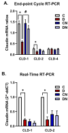
A bar diagram showing the mRNA expression levels as ratios relative to β-actin mRNA of the indicated claudin mRNA isoforms -1, -2, and -4 using (A) the endpoint RT-PCR method and (B) SYBR green RT-PCR-based CT calculations for claudin isoforms -1 and -2 in the LiCl-purified colonic RNA samples from the test conditions: C [non-colitis control], D [DSS-induced colitis], CN [nobiletin-treated non-colitis control], and DN [nobiletin-treated DSS-induced colitis]. The data are the mean ± SE [n = 10]. *, # indicate significance at p < 0.05 between the indicated pair of groups.
Figure 7
Figure 7
A proposed mechanism of the pathogenesis of DSS-induced ulcerative colitis and its reversal by nobiletin treatment in colitis rats. The figure was created using BioRender with permission.

References

    1. Odenwald M.A., Turner J.R. The intestinal epithelial barrier: A therapeutic target? Nat. Rev. Gastroenterol. Hepatol. 2017;14:9–21. doi: 10.1038/nrgastro.2016.169. - DOI - PMC - PubMed
    1. Lechuga S., Ivanov A.I. Disruption of the epithelial barrier during intestinal inflammation: Quest for new molecules and mechanisms. Biochim. Biophys. Acta. 2017;1864:1183–1194. doi: 10.1016/j.bbamcr.2017.03.007. - DOI - PMC - PubMed
    1. Barmeyer C., Fromm M., Schulzke J.D. Active and passive involvement of claudins in the pathophysiology of intestinal inflammatory diseases. Pflügers Arch.-Eur. J. Physiol. 2017;469:15–26. doi: 10.1007/s00424-016-1914-6. - DOI - PubMed
    1. Choi W., Yeruva S., Turner J.R. Contributions of intestinal epithelial barriers to health and disease. Exp. Cell Res. 2017;358:71–77. doi: 10.1016/j.yexcr.2017.03.036. - DOI - PMC - PubMed
    1. Bischoff S.C., Barbara G., Buurman W., Ockhuizen T., Schulzke J.D., Serino M., Tilg H., Watson A., Wells J.M. Intestinal permeability—A new target for disease prevention and therapy. BMC Gastroenterol. 2014;14:189. doi: 10.1186/s12876-014-0189-7. - DOI - PMC - PubMed

LinkOut - more resources