. 2024 Sep 5;14(9):1124.
doi: 10.3390/biom14091124.

Urinary Extracellular Vesicles for Non-Invasive Quantification of Principal Cell Damage in Kidney Transplant Recipients

Affiliations

Urinary Extracellular Vesicles for Non-Invasive Quantification of Principal Cell Damage in Kidney Transplant Recipients

Per Svenningsen et al. Biomolecules. .

Abstract

The objective of the present study was to compare principal cell-specific aquaporin-2 (AQP2) abundances in urinary extracellular vesicles (uEVs) on the first postoperative day in deceased-donor kidney transplant recipients without and with acute kidney injury. We measured uEV markers (CD9 and CD63) and the abundances of proximal tubular sodium-glucose transporter 2, distal tubular sodium/chloride cotransporter, and principal cell-specific aquaporin-2 using Western blotting of urine. uEV-AQP2 levels were normalized to living donor controls. The validation cohort consisted of 82 deceased-donor kidney transplant recipients who had a median age of 50 years (IQR 43 to 57 years). A total of 32% of recipients had acute kidney injury. The median uEV-AQP2 was significantly higher in recipients with acute kidney injury compared to immediate allograft function (2.05; IQR 0.87 to 2.83; vs. 0.81; IQR 0.44 to 1.78; p < 0.01). The Youden index indicated a uEV-AQP2 threshold of 2.00. Stratifying uEV-AQP2 into quartiles showed that recipients with higher uEV-AQP2 levels had higher rates of acute kidney injury (Cochran-Armitage, p = 0.001). The discovery cohort showed elevated CD9, CD63, and uEV-AQP2 levels in urine from recipients with acute kidney injury compared to immediate allograft function. We were able to quantify the damage of principal cells after kidney transplant to predict acute kidney injury using uEV-AQP2.

Keywords: allograft injury; aquaporin-2 abundance; damage of principal cells; exosomes; extracellular vesicles; kidney transplant; urinary extracellular vesicle.


Conflict of interest statement

The authors declare no conflicts of interest. The funders had no role in the design of the study; in the collection, analyses, or interpretation of data; in the writing of the manuscript; or in the decision to publish the results.

Figures

Figure 1
Figure 1
Development of monoclonal antibodies against human aquaporin-2 (AQP2). (A). The monoclonal antibodies were raised against the external epitopes of human AQP2, which are indicated with a rectangle. (B). Screening of individual clones for their signal ratio in control and NDI uEVs. The clones showing the highest signal (black bars) were selected, i.e., clones 19-64-17 and 19-65-1. (C). Signal determination with different monoclonal capture antibodies. Anti-CD9 capture antibodies produced the highest signal compared to anti-CD63 and anti-CD81.
Figure 1
Figure 1
Development of monoclonal antibodies against human aquaporin-2 (AQP2). (A). The monoclonal antibodies were raised against the external epitopes of human AQP2, which are indicated with a rectangle. (B). Screening of individual clones for their signal ratio in control and NDI uEVs. The clones showing the highest signal (black bars) were selected, i.e., clones 19-64-17 and 19-65-1. (C). Signal determination with different monoclonal capture antibodies. Anti-CD9 capture antibodies produced the highest signal compared to anti-CD63 and anti-CD81.
Figure 2
Figure 2
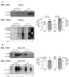
Representative Western blots of the expression of specific markers of extracellular vesicles, tetraspanin CD9 (molecular weight 24 kD), and tetraspanin CD63 (molecular weight 53 kD). Representative Western blots from 10 kidney transplant recipients with immediate allograft function (IGF, 5 samples) and acute kidney injury (5 samples) are shown, where postoperative urine had been obtained on day 1 (upper panels (A)) and day 29 post-transplant (lower panels (B)). Molecular weight markers are indicated. For quantification, densitometry of the Western blots was performed using Fiji 2. Groups were compared using the non-parametric Mann–Whitney test. (Original Western Blot Images see Supplementary Materials).
Figure 3
Figure 3
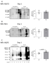
Representative Western blots of the expression of proximal tubular expressed sodium-glucose transporter 2 (SGLT2, upper panel), distal tubular expressed sodium/chloride cotransporter (NCC, middle panel), and principal cell-specific aquaporin-2 (AQP2, lower panel) in urinary extracellular vesicles (uEVs) obtained in post-transplant urine from kidney transplant recipients. Representative Western blots from 10 kidney transplant recipients with immediate allograft function (IGF) and acute kidney injury are shown, where urine was obtained on the first postoperative day. Molecular weight markers are indicated. For quantification, densitometry of the Western blots was performed using Fiji 2. Groups were compared using the non-parametric Mann–Whitney test. (Original Western Blot Images see Supplementary Materials).
Figure 4
Figure 4
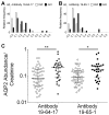
Abundance of principal cell-specific aquaporin-2 (AQP2) in urinary extracellular vesicles (uEVs). Graph showing the distribution of AQP2 abundance in uEVs from kidney transplant recipients with immediate allograft function (open bars) and acute kidney injury (AKI, filled bars). (A). AQP2 antibody 19-64-17. (B). AQP2 antibody 97-65-1. (C). Principal cell-specific AQP2 abundance in uEVs from kidney transplant recipients with immediate allograft function (open circles) and acute kidney injury (filled circles). ** p < 0.01; * p < 0.05 by Mann–Whitney test.
Figure 5
Figure 5
Principal cell-specific AQP2 abundance in urinary extracellular vesicles (uEVs) from deceased donors normalized to living donors with immediate allograft function as controls. (A). AQP2 abundance in uEVs from kidney transplant recipients with immediate allograft function (open circles) and acute kidney injury (AKI, filled circles). Left panel, specific AQP2 antibody 19-64-17; right panel, specific AQP2 antibody 97-65-1. Receiver operating characteristic (ROC) curve for prediction of acute kidney injury from AQP2 abundance in uEVs using antibody 19-64-17 (B) and antibody 97-65-1 (C). Graph showing specificity (open circles) and Youden index (filled circles) of AQP2 abundance in uEVs (fold increase of control) to predict acute kidney injury using antibody 19-64-17 (D) and antibody 97-65-1 (E).
Figure 6
Figure 6
Cochran–Armitage test of trend between quartiles of abundance of aquaporin-2 in urinary extracellular vesicles (uEV-AQP2) and percentage of transplant recipients who experienced acute kidney injury in the validation cohort. The Cochran–Armitage test showed p = 0.002 and p = 0.01 when using 19-64-17 antibodies (A) and 19-65-1 antibodies (B), respectively.

Similar articles

Cited by

References

    1. Johnston O., O’Kelly P., Spencer S., Donohoe J., Walshe J.J., Little D.M., Hickey D., Conlon P.J. Reduced graft function (with or without dialysis) vs immediate graft function--A comparison of long-term renal allograft survival. Nephrol. Dial. Transplant. 2006;21:2270–2274. doi: 10.1093/ndt/gfl103. - DOI - PubMed
    1. Siedlecki A., Irish W., Brennan D.C. Delayed graft function in the kidney transplant. Am. J. Transplant. 2011;11:2279–2296. doi: 10.1111/j.1600-6143.2011.03754.x. - DOI - PMC - PubMed
    1. Aranda-Rivera A.K., Cruz-Gregorio A., Pedraza-Chaverri J., Scholze A. Nrf2 Activation in Chronic Kidney Disease: Promises and Pitfalls. Antioxidants. 2022;11:1112. doi: 10.3390/antiox11061112. - DOI - PMC - PubMed
    1. Rasmussen M., Hansen K.H., Scholze A. Nrf2 Protein Serum Concentration in Human CKD Shows a Biphasic Behavior. Antioxidants. 2023;12:932. doi: 10.3390/antiox12040932. - DOI - PMC - PubMed
    1. Nagarajah S., Rasmussen M., Hoegh S.V., Tepel M. Prospective Study of Long Noncoding RNA, MGAT3-AS1, and Viremia of BK Polyomavirus and Cytomegalovirus in Living Donor Renal Transplant Recipients. Kidney Int. Rep. 2020;5:2218–2227. doi: 10.1016/j.ekir.2020.09.005. - DOI - PMC - PubMed

Grants and funding

LinkOut - more resources