. 2024 Sep 6;14(9):1131.
doi: 10.3390/biom14091131.

An Enhanced Retroviral Vector for Efficient Genetic Manipulation and Selection in Mammalian Cells

Affiliations

An Enhanced Retroviral Vector for Efficient Genetic Manipulation and Selection in Mammalian Cells

Jana Triller et al. Biomolecules. .

Abstract

Introducing genetic material into hard-to-transfect mammalian cell lines and primary cells is often best achieved through retroviral infection. An ideal retroviral vector should offer a compact, selectable, and screenable marker while maximizing transgene delivery capacity. However, a previously published retroviral vector featuring an EGFP/Puromycin fusion protein failed to meet these criteria in our experiments. We encountered issues such as low infection efficiency, weak EGFP fluorescence, and selection against infected cells. To address these shortcomings, we developed a novel retroviral vector based on the Moloney murine leukemia virus. This vector includes a compact bifunctional EGFP and Puromycin resistance cassette connected by a 2A peptide. Our extensively tested vector demonstrated superior EGFP expression, efficient Puromycin selection, and no growth penalty in infected cells compared with the earlier design. These benefits were consistent across multiple mammalian cell types, underscoring the versatility of our vector. In summary, our enhanced retroviral vector offers a robust solution for efficient infection, reliable detection, and effective selection in mammalian cells. Its improved performance and compact design make it an ideal choice for a wide range of applications involving precise genetic manipulation and characterization in cell-based studies.

Keywords: B cells; EGFP; FACS; Puromycin; T2A; fusion protein; infection; linker; retrovirus.


Conflict of interest statement

The authors declare no conflicts of interest. The funders had no role in the design of the study; in the collection, analyses, or interpretation of data; in the writing of the manuscript; or in the decision to publish the results.

Figures

Figure 1
Figure 1
Flow cytometric analyses for EGFP- and TagBFP fluorescence of 38B9 cells infected with different retroviral supernatants. 38B9 cells were (A) mock infected or infected with different retroviral supernatants derived from (B) pBMN-I-GFP, (C) pBMN-I-TagBFP, (D) pBMN-I-EGFP/Puro, and (E) pBMN-I-EGFP/Puro long and incubated for one day before flow cytometric analysis to determine infection efficiencies. A detailed gating and analysis strategy is shown in Supplemental Figure S4. Cells (COUNT) are shown in histogram plots for EGFP- [FI (EGFP), gate “I”, upper row] or BFP- [FI (BFP), gate “H”, bottom row] fluorescence intensities. Percentages of gated cells are shown in the graphs.
Figure 2
Figure 2
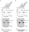
Western blot analysis of EGFP proteins in wild-type- and infected 38B9- and NIH3T3 cells. Cell lysates from 1 × 10⁶ uninfected and unselected (A) 38B9- or (B) NIH3T3 cells (“WT”) or from 1 × 10⁶ (A) 38B9- or (B) NIH3T3 cells infected with retroviral supernatant from the respective vectors pBMN-I-EGFP-T2A-Puro (“EGFP-T2A-Puro”), pBMN-I-EGFP/Puro (“EGFP/Puro”), pBMN-I-EGFP/Puro long (“EGFP/Puro long”), pBMN-I-EGFP/Puro Myc long (“EGFP/Puro Myc long”) or pBMN-I-EGFP-T2A-Puro Myc (“EGFP-T2A-Puro Myc”) and Puromycin-selected for 5 days were reduced, separated by 13.5% SDS-PAGE, and transferred to a nitrocellulose membrane. The membrane was blocked, stained with monoclonal mouse anti-GFP antibodies, and developed with an appropriate HRP-conjugated secondary antibody using the ECL method for a short (upper panel) or prolonged exposure (middle panel). The loading of same cell equivalents was assessed with polyclonal rabbit antibodies against beta-actin (bottom panel). Dashed arrows in the beta-actin blots indicate signals from previous probing with the GFP antibody. Original images can be found in Supplemental Figures S9 and S10.
Figure 3
Figure 3
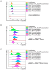
Flow cytometric analyses for EGFP fluorescence in 38B9 cells infected with EGFP/Puro cassette-containing retroviral supernatants and subsequent Puromycin selection. 38B9 cells were infected with retroviral supernatants derived from (A) mock-, (B) pBMN-I-EGFP/Puro-, or (C) pBMN-I-EGFP/Puro long-transfected Platinum-E cells. Puromycin was added at 5 µg/mL 24 h after infection. EGFP fluorescence intensities to determine infection efficiency were measured by flow cytometry on the day of infection, one day after infection, and every other day for 5 days. Data acquisition and gating strategy were identical to those described in Figure 1. EGFP fluorescence intensities of FSC/SSC-gated single cells are presented as overlay histograms, and relative cell numbers of each measurement are normalized and presented as %Max.
Figure 4
Figure 4
Flow cytometric analyses for EGFP fluorescence in 38B9 cells infected with EGFP-T2A-Puro cassette-containing retroviral supernatants and subsequent Puromycin selection. 38B9 cells were infected with retroviral supernatants derived from (A) mock-, (B) pBMN-I-EGFP-T2A-Puro-, or (C) pBMN-I-EGFP-T2A-Puro Myc-transfected Platinum-E cells. Puromycin was added at 5 µg/mL 24 h after infection. EGFP fluorescence intensities to determine infection efficiency were measured by flow cytometry on the day of infection, one day after infection, and every other day for 5 days. Data acquisition and gating strategy were identical to those described in Figure 1. Data of mock infection are identical to those in Figure 3. EGFP fluorescence intensities of FSC/SSC-gated single cells are presented as overlay histograms, and relative cell numbers of each measurement are normalized and presented as %Max.

References

    1. Banerjee P., Crawford L., Samuelson E., Feuer G. Hematopoietic stem cells and retroviral infection. Retrovirology. 2010;7:8. doi: 10.1186/1742-4690-7-8. - DOI - PMC - PubMed
    1. Saez-Cirion A., Manel N. Immune Responses to Retroviruses. Annu. Rev. Immunol. 2018;36:193–220. doi: 10.1146/annurev-immunol-051116-052155. - DOI - PubMed
    1. Vara J., Perez-Gonzalez J.A., Jimenez A. Biosynthesis of puromycin by Streptomyces alboniger: Characterization of puro-mycin N-acetyltransferase. Biochemistry. 1985;24:8074–8081. doi: 10.1021/bi00348a036. - DOI - PubMed
    1. Miller A.D. Development and applications of retroviral vectors. In: Coffin J.M., Hughes S.H., Varmus H.E., editors. Retroviruses. Cold Spring Harbor; New York, NY, USA: 1997. - PubMed
    1. Martinez-Salas E., Francisco-Velilla R., Fernandez-Chamorro J., Embarek A.M. Insights into Structural and Mechanistic Features of Viral IRES Elements. Front. Microbiol. 2017;8:2629. doi: 10.3389/fmicb.2017.02629. - DOI - PMC - PubMed

Substances

LinkOut - more resources