. 2024 Sep 20;14(9):1187.
doi: 10.3390/biom14091187.

Improving Circulation Half-Life of Therapeutic Candidate N-TIMP2 by Unfolded Peptide Extension

Affiliations

Improving Circulation Half-Life of Therapeutic Candidate N-TIMP2 by Unfolded Peptide Extension

Jason Shirian et al. Biomolecules. .

Abstract

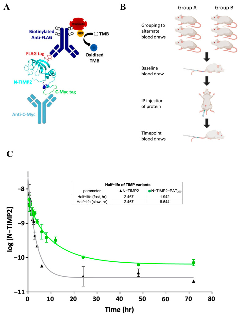
Matrix metalloproteinases (MMPs) are significant drivers of many diseases, including cancer, and are established targets for drug development. Tissue inhibitors of metalloproteinases (TIMPs) are endogenous MMP inhibitors and are being pursued for the development of anti-MMP therapeutics. TIMPs possess many attractive properties for drug candidates, such as complete MMP inhibition, low toxicity, low immunogenicity, and high tissue permeability. However, a major challenge with TIMPs is their rapid clearance from the bloodstream due to their small size. This study explores a method for extending the plasma half-life of the N-terminal domain of TIMP2 (N-TIMP2) by appending it with a long, intrinsically unfolded tail containing Pro, Ala, and Thr (PATylation). We designed and produced two PATylated N-TIMP2 constructs with tail lengths of 100 and 200 amino acids (N-TIMP2-PAT100 and N-TIMP2-PAT200). Both constructs demonstrated higher apparent molecular weights and retained high inhibitory activity against MMP-9. N-TIMP2-PAT200 significantly increased plasma half-life in mice compared to the non-PATylated variant, enhancing its therapeutic potential. PATylation offers distinct advantages for half-life extension, such as fully genetic encoding, monodispersion, and biodegradability. It can be easily applied to N-TIMP2 variants engineered for high affinity and selectivity toward individual MMPs, creating promising candidates for drug development against MMP-related diseases.

Keywords: PASylation; PATylation; TIMP; half-life extension; matrix metalloprotein inhibitors; therapeutic protein.


Conflict of interest statement

The authors declare no conflicts of interest.

Figures

Figure 1
Figure 1
Preparation of N-TIMP2 tagged and PATylated constructs. (A) Schematic representation of expressed constructs of N-TIMP2 (top) and PATylated constructs (bottom). The N-TIMP2 domain is shown in cyan, the internal FLAG-tag in red, c-myc tag in green and 6xHis tag in blue. (B) Ten model decoys of N-TIMP2-PAT100 fused to a 100 amino acid PAT tail as modeled by AlfaFold. N-TIMP2 is shown in cyan, PAT fusion in orange, and FLAG-tag in red. In comparison, the actual structure of N-TIMP2 is shown in green (PDB ID 1BR9). The structures of N-TIMP2-PAT200 looked generally similar to those of N-TIMP2-PAT100.
Figure 2
Figure 2
Characterization of N-TIMP2 and the PATylated variants. (A) SDS-PAGE under nonreducing conditions followed by silver staining of N-TIMP2 and two preps of N-TIMP2-PAT200 (left) and N-TIMP2-PAT100 (right). (B) Dynamic light scattering (DLS) validation of increased hydrodynamic radius in PATylated N-TIMP2. Superposed particle size distributions of N-TIMP2 (black) with peak at 2.806 nm, N-TIMP2-PAT100 (red) with peak at 4.253 nm, and N-TIMP2-PAT200 (blue) with peak at 5.612 nm. (C) CD spectra of the three constructs: N-TIMP2 (blue), N-TIMP2-PAT100 (orange), and N-TIMP2-PAT200 (green). Each curve is an average of 5 runs. (D) Inhibitory activity of the N-TIMP2 (blue), N-TIMP2-PAT100 (orange), and N-TIMP2-PAT200 (green) against MMP-9. Fraction of MMP-9 activity is plotted vs. concentration of added N-TIMP2-based inhibitor. The data were fitted to Equation (1) to obtain Kiapp.
Figure 3
Figure 3
Pharmacokinetic study in mice demonstrates significant extension of plasma half-life for N-TIMP2-PAT200. (A) A schematic diagram of the ELISA setup for the detection of recombinant N-TIMP2-based constructs from plasma. An anti-C-myc tag antibody on the surface of a MaxiSorp™ plate is used for capturing the N-TIMP2 constructs from plasma. The protein is detected by a biotinylated anti-FLAG-tag antibody, which binds to HRP-conjugated streptavidin that in turn catalyzes the oxidation of the TMB substrate to the measurable blue colored product. The blue, green, and red regions highlighted on the N-TIMP2 protein, respectively, represent the 6xHIS tag, C-myc tag, and FLAG tag. (B) Schematic diagram of pharmacokinetic study design, in which each N-TIMP2 variant was tested using 6 mice grouped into two subcohorts of 3 mice each, which were bled at alternating time points following the initial baseline bleed and IP injection of N-TIMP proteins. (C) Elimination half-life of N-TIMP2 black triangles versus N-TIMP2-PAT200 (green circles). Plot of average ± SEM (n = 3) of log10 N-TIMP2 concentration vs. time, modeled with a 2-phase decay with least-squares nonlinear fit for half-life determination. Statistical significance assessed via a sum-of-squares F test where H0: Kslow N-TIMP2-PAT200 = Kslow N-TIMP2; Ha: Kslow N-TIMP2-PAT200 ≠ Kslow N-TIMP2. A = 0.05, p < 0.0001.

Update of

References

    1. Vandenbroucke R.E., Libert C. Is there new hope for therapeutic matrix metalloproteinase inhibition? Nat. Rev. Drug Discov. 2014;13:904–927. doi: 10.1038/nrd4390. - DOI - PubMed
    1. Radisky E.S., Coban M. Enzymes|Matrix Metalloproteinases. In: Jez J., editor. Encyclopedia of Biological Chemistry. 3rd ed. Elsevier; Amsterdam, The Netherlands: 2021. pp. 336–353.
    1. Cabral-Pacheco G.A., Garza-Veloz I., Castruita-De la Rosa C., Ramirez-Acuna J.M., Perez-Romero B.A., Guerrero-Rodriguez J.F. The Roles of Matrix Metalloproteinases and Their Inhibitors in Human Diseases. Int. J. Mol. Sci. 2020;21:9739. doi: 10.3390/ijms21249739. - DOI - PMC - PubMed
    1. Mason S.D., Joyce J.A. Proteolytic networks in cancer. Trends Cell Biol. 2011;21:228–237. doi: 10.1016/j.tcb.2010.12.002. - DOI - PMC - PubMed
    1. Radisky E.S. Extracellular proteolysis in cancer: Proteases, substrates, and mechanisms in tumor progression and metastasis. J. Biol. Chem. 2024;300:107347. doi: 10.1016/j.jbc.2024.107347. - DOI - PMC - PubMed

MeSH terms

Substances

LinkOut - more resources