Review
. 2024 Sep 21;14(9):1192.
doi: 10.3390/biom14091192.

Significance of Necroptosis in Cartilage Degeneration

Affiliations
Review

Significance of Necroptosis in Cartilage Degeneration

Md Abdul Khaleque et al. Biomolecules. .

Abstract

Cartilage, a critical tissue for joint function, often degenerates due to osteoarthritis (OA), rheumatoid arthritis (RA), and trauma. Recent research underscores necroptosis, a regulated form of necrosis, as a key player in cartilage degradation. Unlike apoptosis, necroptosis triggers robust inflammatory responses, exacerbating tissue damage. Key mediators such as receptor-interacting serine/threonine-protein kinase-1 (RIPK1), receptor-interacting serine/threonine-protein kinase-3(RIPK3), and mixed lineage kinase domain-like (MLKL) are pivotal in this process. Studies reveal necroptosis contributes significantly to OA and RA pathophysiology, where elevated RIPK3 and associated proteins drive cartilage degradation. Targeting necroptotic pathways shows promise; inhibitors like Necrostatin-1 (Nec-1), GSK'872, and Necrosulfonamide (NSA) reduce necroptotic cell death, offering potential therapeutic avenues. Additionally, autophagy's role in mitigating necroptosis-induced damage highlights the need for comprehensive strategies addressing multiple pathways. Despite these insights, further research is essential to fully understand necroptosis' mechanisms and develop effective treatments. This review synthesizes current knowledge on necroptosis in cartilage degeneration, aiming to inform novel therapeutic approaches for OA, RA, and trauma.

Keywords: OA; RA; cartilage degeneration; necroptosis; trauma.


Conflict of interest statement

The authors declare no conflicts of interest.

Figures
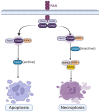
Figure 1
Figure 1
The graphical presentation of common initiation executes significantly different pathways of apoptosis and necroptosis.
Figure 2
Figure 2
A comparison between healthy and degenerated cartilage in the knee joints.
Figure 3
Figure 3
Typical molecular mechanisms of necroptosis: Necroptosis-mediated cell death is initiated by the activation of death receptors such as the TNF receptor, FAS, Toll-like receptor, or interferon receptor. These receptors activate RIPK1 or other RIP homologous interaction motif (RHIM) domain-containing proteins, which then interact with RIPK3 to form the necrosome complex. RIPK3 is activated through phosphorylation and subsequently phosphorylates MLKL. Phosphorylated MLKL oligomerizes and moves to the plasma membrane, triggering necroptosis. RIPK1, RIPK3, and MLKL are the core components of TNF-induced necroptosis. Additionally, RIPK3 can activate CaMK II, leading to the opening of the mPTP and necroptosis in cardiomyocytes. RIPK3 can also be activated by other RHIM domain-containing proteins like TRIF and DAI, expanding the mechanisms of RIPK3.
Figure 4
Figure 4
Categorization of arthritis.
Figure 5
Figure 5
Molecular mechanisms of necroptosis in OA and TMJOA: In TMJOA, TNFα induces Syndecan 4 (SDC4), which amplifies TNFα signaling and triggers necroptosis, releasing cartilage-degrading enzymes and intensifying inflammation. Inhibiting RIPK3, pMLKL, and SDC4 protects cartilage and reduces inflammation. BMP7 induces necroptosis through RIP1, with BMP7 silencing reducing RIPK1-induced necroptosis and restoring ECM gene expression. High RIPK3 expression accelerates cartilage degradation, while RIPK3 inhibition by AZ-628 mitigates OA progression. TRADD inhibition with ICCB-19 blocks the RIPK1-TAK1 pathway, reducing inflammation and necroptosis. PLCγ1 inhibition, combined with apoptosis and necroptosis blockers, enhances cartilage matrix synthesis. RIPK1 knockdown disrupts the TRIF/MyD88-RIPK1-TRAF2 pathway, alleviating OA. AZD8330 activates cIAP1, inhibiting RIPK1-associated necrosis, and preserving cartilage.
Figure 6
Figure 6
Molecular mechanisms of necroptosis in RA: Nec-1 and amiloride inhibit necroptosis in RA chondrocytes by targeting the RIP1/RIP3/p-MLKL pathway, with ASIC1a-mediated upregulation reversible by PcTx-1 or Nec-1. IFN-γ mitigates necroptosis and inflammation by reducing MLKL and modulating inflammatory responses, despite its proinflammatory role. KW2449 ameliorates collagen-induced arthritis by inhibiting RIPK1-dependent necroptosis, reducing RIPK1 and MLKL levels. Irisin reduces necroptotic signaling and inflammation via the NF-kB and Nrf2/HO-1 pathways, downregulating TNF-α, MCP1, and HMGB1, promoting chondrocyte recovery.
Figure 7
Figure 7
Molecular mechanisms of necroptosis in trauma: Nec-1, a RIPK1 inhibitor, surpasses zVAD in protecting against trauma-induced necroptosis by reducing MLKL expression and PGE2 production. ROS are crucial in RIPK1-mediated necroptosis, which is more prominent in late-stage OA. Necroptosis markers RIPK3 and MLKL in OA cartilage are linked to PGE2 and NO release, and necrostatin-1 inhibits post-trauma necroptosis. In TMJOA, RIP1 inhibition reduces apoptosis and necroptosis. Mechanical stress induces necroptosis in chondrocytes, with Nec-1 and Z-VAD reducing TNF-α-induced ROS and necroptosis. D469del-COMP retention triggers necroptosis via ER stress, oxidative stress, and DNA damage.

References

    1. Sophia Fox A.J., Bedi A., Rodeo S.A. The Basic Science of Articular Cartilage: Structure, Composition, and Function. Sports Health. 2009;1:461–468. doi: 10.1177/1941738109350438. - DOI - PMC - PubMed
    1. Marc C.H., editor. Rheumatoid Arthritis. Mosby; Philadelphia, PA, USA: 2009. CHAPTER 8G—Chondrocytes: Pathogenesis of Rheumatoid Arthritis; pp. 151–162.
    1. Buckwalter J.A., Mankin H.J., Grodzinsky A.J. Articular cartilage and osteoarthritis. Instr. Course Lect. -Am. Acad. Orthop. Surg. 2005;54:465. - PubMed
    1. Stockwell R.A. Biology of cartilage cells. 1979: CUP Archive.Neogi, T. The epidemiology and impact of pain in osteoarthritis. Osteoarthr. Cartil. 2013;21:1145–1153. doi: 10.1016/j.joca.2013.03.018. - DOI - PMC - PubMed
    1. Neogi T. The epidemiology and impact of pain in osteoarthritis. Osteoarthr. Cartil. 2013;21:1145–1153. doi: 10.1016/j.joca.2013.03.018. - DOI - PMC - PubMed

LinkOut - more resources