. 2024 Sep 15;13(9):889.
doi: 10.3390/antibiotics13090889.

Three in One with Dual-Functional Hydrogel of Lactoferrin/NZ2114/LMSH Promoting Staphylococcus aureus-Infected Wound Healing

Affiliations

Three in One with Dual-Functional Hydrogel of Lactoferrin/NZ2114/LMSH Promoting Staphylococcus aureus-Infected Wound Healing

Kun Zhang et al. Antibiotics (Basel). .

Abstract

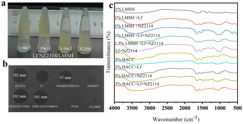
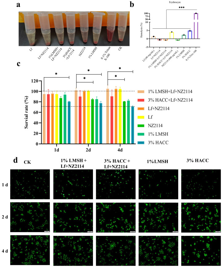
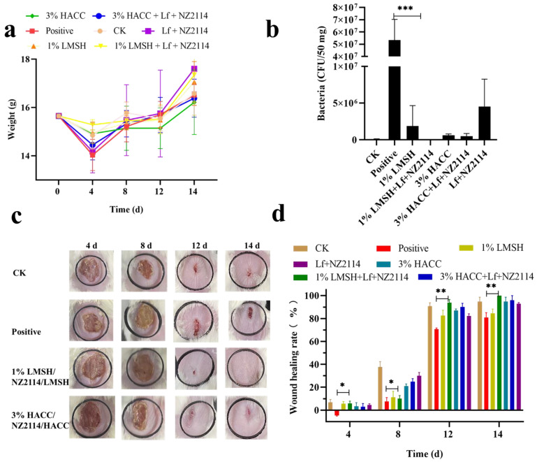
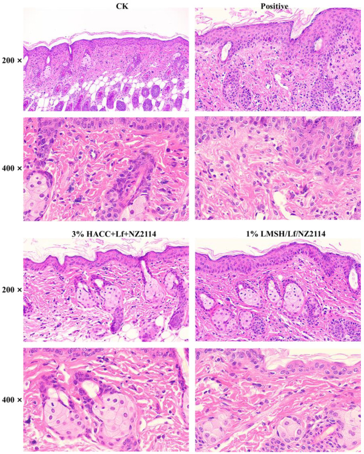
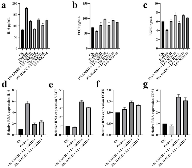
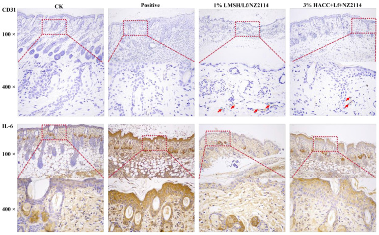
Wound infections caused by Staphylococcus aureus often result in localized suppurative lesions that severely impede the healing process, so it is urgent to develop a dress with efficient antimicrobial and pro-healing functions. In this study, the bifunctional injectable hydrogel lactoferrin (Lf)/NZ2114/lithium magnesium silicate hydrogel (LMSH) was first successfully prepared through the electrostatic interaction method. The physical, biological, and efficacy properties are systematically analyzed with good shear-thinning capacity and biocompatibility. More importantly, it inhibits infection and promotes wound healing in a mouse wound infection model after 14 d treatment, and the bactericidal rate and healing rate were over 99.92% and nearly 100%, respectively. Meanwhile, the massive reduction of inflammatory cells, restoration of tissue structure, and angiogenesis in mice showed the anti-inflammatory and pro-healing properties of the hydrogel. The healed wounds showed thickening with more hair follicles and glands, suggesting that the hydrogel Lf/NZ2114/LMSH (Three in One) could be a better dressing candidate for the treatment of S. aureus-induced wound infections.

Keywords: Lf/NZ2114/LMSH (Three in One); Staphylococcus aureus infection; injectable hydrogel; would dressing.


Conflict of interest statement

The authors declare no competing interests.

Figures

Figure 1
Figure 1
The photograph, antimicrobial activity and FT–IR spectrum of the injectable hydrogel Lf/NZ2114/LMSH. (a) The photograph of the synthetic injectable hydrogel; (b) Inhibition zone of NZ2114, Lf, 1% LMSH/Lf/NZ2114, 3% HACC/Lf/NZ2114, 3% SA, 1% LMSH; (c) The FT–IR spectra of 1% LMSH, 1% LMSH + Lf, 1% LMSH + NZ2114, 1% LMSH + Lf + NZ2114, 1.5% LMSH + Lf + NZ2114, 3% HACC, 3% HACC + Lf, 3% HACC + NZ2114, 3% HACC + Lf + NZ2114.
Figure 2
Figure 2
The morphology of the synthetic injectable hydrogel. The SEM image of 0.5% LMSH + NZ2114, 1% LMSH + Lf + NZ2114, 1% LMSH + Lf + NZ2114, 3% HACC + Lf + NZ2114, 1% LMSH, 3% HACC, Lf + NZ2114.
Figure 3
Figure 3
The viscosity, modulus, and bactericidal properties of different hydrogel samples. (a) The viscosity of 0.5% Lf/NZ2114/LMSH and 1% Lf/NZ2114/LMSH during sheer increase from 0.01 to 100 s−1; (b) The viscosity of 3% Lf/NZ2114/HACC during sheer increase from 0.01 to 100 s−1; (c) The storage modulus (G′) and loss modulus (G″) of 0.5% Lf/NZ2114/LMSH, 1% Lf/NZ2114/LMSH, and 3% Lf/NZ2114/HACC during strain increase from 0.1% to 1000% at the frequency of 1 Hz; (d,e) The bactericidal rate of 1% Lf/NZ2114/LMSH, 3% Lf/NZ2114/HACC, Lf + NZ2114, Lf, NZ2114, 1% LMSH, 3% HACC against S. aureus CVCC 546 (n = 3). These (−2, −3, −4, −5) are the number of dilutions. (* p < 0.05, *** p < 0.001).
Figure 4
Figure 4
The biocompatibility of different hydrogel samples. (a,b) The images and hemolysis rate of Lf, Lf + NZ2114, 1% Lf/NZ2114/LMSH, 3% Lf/NZ2114/HACC, 1% LMSH, 3% HACC, 0.1% Trix-100; (c) Cytotoxicity of HACAT cells co-cultured with Lf, Lf + NZ2114, 1% Lf/NZ2114/LMSH, 3% Lf/NZ2114/HACC, 1% LMSH, 3% HACC. Samples at 1, 2, and 4 days, n = 3; (d) The images of calcein–AM/PI double staining of the HACAT cells that were incubated with 1% Lf/NZ2114/LMSH, 3% Lf/NZ2114/HACC, 1% LMSH, 3% HACC for 1, 2 and 4 days. (Scale bar = 100 μm). * p < 0.05, *** p < 0.001.
Figure 5
Figure 5
The body weight, skin load, and wound diagram of mice (n = 3). (a) The weight of mice untreated and treated with Lf + NZ2114, 1% Lf/NZ2114/LMSH, 3% Lf/NZ2114/HACC, 1% LMSH, 3% HACC for 0–14 days; (b) The bacteria of skin untreated and treated with Lf + NZ2114, 1% Lf/NZ2114/LMSH, 3% Lf/NZ2114/HACC, 1% LMSH, 3% HACC samples for 4 d; (c) The macroscopic images of wounds untreated and treated with 1% Lf/NZ2114/LMSH, 3% Lf/NZ2114/HACC samples for 4, 8, 12 and 14 d; (d) The wound healing rate of mice untreated and treated with Lf + NZ2114, 1% Lf/NZ2114/LMSH, 3% Lf/NZ2114/HACC, 1% LMSH, 3% HACC samples for 4, 8, 12, and 14 d. * p < 0.05, ** p < 0.01, *** p < 0.001.
Figure 6
Figure 6
HE staining of the wounds without any treatment or treated with 1% Lf/NZ2114/LMSH and 3% Lf/NZ2114/HACC samples for 14 days.
Figure 7
Figure 7
The cytokine secretion without any treatment or treated with 1% Lf/NZ2114/LMSH and 3% Lf/NZ2114/HACC samples for 14 days. (ac) The expressions of IL-6, VEGF and EGFR were measured by ELISA kit. (dg) The expressions of IL-6, VEGF, EGFR, and CD31 were measured at RNA level by qPCR.
Figure 8
Figure 8
The immunohistochemistry of without any treatment or treated with 1% Lf/NZ2114/LMSH and 3% Lf/NZ2114/HACC samples for 14 days. CD31 staining of the wounds without any treatment or treated with different hydrogel samples for 14 days. IL-6 staining of the wounds for 14 days. (Red arrows are CD31 binding sites).

References

    1. Serra R., Grande R., Butrico L., Rossi A., Settimio U.F., Caroleo B., Amato B., Gallelli L., De Franciscis S. Chronic wound infections: The role of Pseudomonas aeruginosa and Staphylococcus aureus. Expert Rev. Anti-Infect. Ther. 2015;13:605–613. doi: 10.1586/14787210.2015.1023291. - DOI - PubMed
    1. Roy S., Santra S., Das A., Dixith S., Sinha M., Ghatak S., Ghosh N., Banerjee P., Khanna S., Mathew-Steiner S. Staphylococcus aureus biofilm infection compromises wound healing by causing deficiencies in granulation tissue collagen. Ann. Surg. 2020;271:1174–1185. doi: 10.1097/SLA.0000000000003053. - DOI - PMC - PubMed
    1. Huang Y., Yang N., Teng D., Mao R., Hao Y., Ma X., Wei L., Wang J. Antibacterial peptide nz2114-loaded hydrogel accelerates Staphylococcus aureus-infected wound healing. Appl. Microbiol. Biotechnol. 2022;106:3639–3656. doi: 10.1007/s00253-022-11943-w. - DOI - PubMed
    1. Martin P., Nunan R. Cellular and molecular mechanisms of repair in acute and chronic wound healing. Br. J. Dermatol. 2015;173:370–378. doi: 10.1111/bjd.13954. - DOI - PMC - PubMed
    1. Huang C., Leavitt T., Bayer L.R., Orgill D.P. Effect of negative pressure wound therapy on wound healing. Curr. Probl. Surg. 2014;51:301–331. doi: 10.1067/j.cpsurg.2014.04.001. - DOI - PubMed

LinkOut - more resources