. 2024 Sep 15;16(18):3167.
doi: 10.3390/cancers16183167.

GABA(A) Receptor Activation Drives GABARAP-Nix Mediated Autophagy to Radiation-Sensitize Primary and Brain-Metastatic Lung Adenocarcinoma Tumors

Affiliations

GABA(A) Receptor Activation Drives GABARAP-Nix Mediated Autophagy to Radiation-Sensitize Primary and Brain-Metastatic Lung Adenocarcinoma Tumors

Debanjan Bhattacharya et al. Cancers (Basel). .

Abstract

In non-small cell lung cancer (NSCLC) treatment, radiotherapy responses are not durable and toxicity limits therapy. We find that AM-101, a synthetic benzodiazepine activator of GABA(A) receptor, impairs the viability and clonogenicity of both primary and brain-metastatic NSCLC cells. Employing a human-relevant ex vivo 'chip', AM-101 is as efficacious as docetaxel, a chemotherapeutic used with radiotherapy for advanced-stage NSCLC. In vivo, AM-101 potentiates radiation, including conferring a significant survival benefit to mice bearing NSCLC intracranial tumors generated using a patient-derived metastatic line. GABA(A) receptor activation stimulates a selective-autophagic response via the multimerization of GABA(A) receptor-associated protein, GABARAP, the stabilization of mitochondrial receptor Nix, and the utilization of ubiquitin-binding protein p62. A high-affinity peptide disrupting Nix binding to GABARAP inhibits AM-101 cytotoxicity. This supports a model of GABA(A) receptor activation driving a GABARAP-Nix multimerization axis that triggers autophagy. In patients receiving radiotherapy, GABA(A) receptor activation may improve tumor control while allowing radiation dose de-intensification to reduce toxicity.

Keywords: GABA receptor; GABARAP; Nix; autophagy; benzodiazepine; brain metastasis; lung cancer; p62; radiotherapy.


Conflict of interest statement

The authors declare no conflicts of interest.

Figures

Figure 1
Figure 1
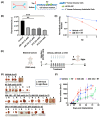
Activation of GABA(A) receptors triggers cell depolarization and death. (A) Type-A GABA (GABA(A)) receptors are ligand-gated chloride anion channels. Left, GABA(A) receptors move chloride anions (Cl) out of the cell during embryonic stages of development but into the cell in mature or developed stages and are thereby depolarizing or hyperpolarizing, respectively. Right, GABA(A) receptors form hetero-pentameric structures with an α2β2γ1 stoichiometry. Two molecules of GABA (purple spheres) bind at the α-β interfaces to ‘activate’ receptor function (chloride anion transport). Commonly, one molecule of benzodiazepine (red sphere) binds at the α-γ interface to enhance flow of chloride anions. Created with BioRender.com (B) Left, GABRA5 or α5 protein in NSCLC patient-derived primary cell lines representing three histological subtypes (adenocarcinoma, A549, H1792; squamous cell, H1703; large cell, H460). Right, GABRA5 or α5 protein expression in patient-derived lung adenocarcinoma brain-metastatic cell line (UW-lung-16), and primary human lung adenocarcinoma cell line (H1792). The presence of protein confirmed by Western blotting of SDS (4–15% gradient) gels. GAPDH is used as a loading control. Cropped gel lanes from original blots, see Figure S8. (C) NSCLC primary (left) and brain-metastatic (right) patient tumor tissue from the same patient (or matched) stains for GABRA5 or α5 protein, as shown by immunohistochemistry staining at 30× magnification. Arrows show GABRA5 staining in large tumor cells within primary and brain-metastatic lung adenocarcinoma tissue sections. (D) AM-101 (QH-II-066) (274.32 g/mol) is a benzodiazepine analog (left). A representative single cell patch clamp electrophysiology trace of patient-derived adenocarcinoma lung cell line H1792 (right). Cells are responsive to GABA or electro-physiologically functional. Perfusion of cells with AM-101 plus GABA elicits an enhanced response, indicating that GABA(A) receptors are benzodiazepine-responsive or ‘activated’. Representative raw current trace recording with the following parameters: GABA, 1 µM; AM-101, 4 µM. (E) Lung adenocarcinoma (H1792) cells incubated with AM-101 are depolarized, as assessed by the TMRE assay and Fluorescence-Activated Cell Sorting (FACS) analysis. Shown is the degrees of depolarization relative to DMSO-alone treatment and FCCP, which provide negative and positive controls in this experiment, respectively. Parameters: AM-101, 2 µM; FCCP, 10 µM. (F) Half-maximal inhibitory concentration (IC50) values of patient-derived lung primary and brain-metastatic lines representative of three histological lung cancer subtypes, as measured using a viability (MTS) assay and AM-101. Indicated is the KRAS and TP53 mutational status of lines, where M represents mutant, and WT represents wild-type.
Figure 2
Figure 2
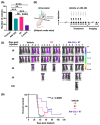
GABA(A) receptor activation potentiates radiation. (A) Illustration of a human-relevant ex vivo ‘chip’ employed to test AM-101 and docetaxel (DTX) efficacy. Lung adenocarcinoma cancer cells (H1792-GFP, green) can be co-cultured with primary human alveolar and pulmonary endothelial cells and exposed to air (air–liquid interface) on-chip. Cancer cells form clusters that grow and spread through the epithelial compartment of the chip over time (Figure S3). (B) Testing of AM-101 and DTX ex vivo or ‘on-chip’ reveals that AM-101 is as cytotoxic as DTX but at a significantly lower concentration. The chip is a 3-D ex vivo model, and, to achieve the cytotoxicity that AM-101 generates in 5 μM concentration, DTX is required in 10 mM concentrations. To determine p-values between two groups, one-way ANOVA with Tukey’s multiple comparisons test was performed. ** p < 0.001 and *** p < 0.0001. Images acquired from chips were subjected to background signal removal and analysis using Fiji (Image J). To generate bar graphs, acquired images were evaluated through background subtraction and signal thresholding. Subsequently, particle analysis was performed using a Fiji plugin to estimate the number of GFP+ cells per field of view under each testing condition. (C) A clonogenic assay was employed to examine the radio-sensitizing effect of AM-101 in H1792 cells. The survival curves showing surviving fraction of H1792 cells following radiation exposure at two separate doses with and without AM-101. Cell cultures were treated with either AM-101 (2.5 μM) combined with two separate doses of radiation (3 Gy and 6 Gy) versus DMSO (vehicle) and two separate doses of radiation (3 Gy and 6 Gy). H1792 cells in culture were treated with AM-101 (2.5 μM) or DMSO (vehicle) 1 h before radiation and maintained in the medium after irradiation. According to the experimental design the media containing AM-101 or DMSO in all groups was replaced with fresh media 72 h after treatment. Colony-forming efficiency was determined 14 days later, and survival curves were generated. The vehicle in this experiment is DMSO, since DMSO is used as the solvent to solubilize AM-101. (D) Schematic of the efficacy experiment in H1792 subcutaneous heterotopic bilateral xenograft tumors generated in NSG mice. Mice in vehicle or drug treatment groups received i.p., vehicle, AM-101 (2.5 mg/kg), or DTX (8 mg/kg), on day 36 post-implantation and then six injections once per day. Mice in radiation (RT) or combo groups received a single fraction of radiation (5 Gy) to left flank only at 2 h before vehicle or drug on the first day of treatment. (E) At experimental endpoint, tumors from left (L) and right (R) flanks of each mouse were resected. H1792 subcutaneous xenograft tumor growth in NSG mice from different treatment groups: vehicle, radiation (RT), AM-101 ± RT, DTX ± RT. Number of mice per treatment group: n = 6 for vehicle, n = 4 for RT; n = 7 for AM-101 and n = 7 for AM-101 + RT, n = 5 for DTX + RT and n = 6 for DTX. (F) Tumor volume of left and right flank tumors was measured over time using Vernier calipers. The tumor growth delay curves show the tumor volumes of mice treated with a vehicle, radiation (RT), AM-101, and AM-101 plus RT. Each point on the curve represents the mean tumor volume after treatment, with error bars indicating the standard error (SE). Statistical significance is indicated by p < 0.001.
Figure 3
Figure 3
GABA(A) receptor activation increases survival of mice bearing lung brain-metastatic tumors. (A) Bar graphs representing the number of colonies generated from clonogenic assay to determine the radiosensitizing effect of AM-101 when combined with radiation treatment (RT) versus AM-101 (2.5 μM) or RT (3 Gy) alone in patient-derived brain-metastatic UW-lung-16 cells. The combination of AM-101 plus RT (combo group) imparts the most significant impact on colony formation, a three-fold suppression of colony numbers than control and two-fold suppression of colony numbers than AM-101 and RT applied alone. Control is DMSO treated, as DMSO is the diluent of AM-101. Data are represented as mean ± S.E. One-way ANOVA with Tukey’s multiple comparisons test was performed to determine the p-values between two treatment groups. The one-way ANOVA p < 0.0001. Based on Tukey’s multiple comparisons test, Control vs. AM-101, *** p = 0.0001; Control vs. RT ** p = 0.0019; Control vs. Combo **** p < 0.0001; AM-101 vs. RT, ns (not significant), p = 0.308; AM-101 vs. Combo *** p = 0.0001; RT vs. Combo **** p < 0.0001. (B) Schematic of efficacy experiment in intracranial xenograft tumors generated in athymic nude mice. Mice received a stereotaxic intracranial injection of cells (UW-lung-16) from a brain lesion of a patient with lung cancer. Mice (n = 21) were separated into three treatment groups (n = 7 per group). Ten days post-injection, mice received (1) vehicle, an i.p. injection of formulation; (2) radiation (RT), 2.5 Gy dose/day to the whole mouse brain for 5 consecutive days; (3) AM-101 plus RT, i.p. injection of formulated AM-101 (5 mg/kg) and RT (2.5 Gy dose/day/mouse for 5 days) to the whole mouse brain using a XenX irradiator (Xstahl Ltd.) and the supplied mouse gantry. The day of intracranial injection of tumor cells was assigned as day zero. Tumors were followed by bioluminescent imaging (BLI) over time. (C) BLI study of mice with brain metastatic lung tumors and treated with vehicle, radiation (RT), or RT plus AM-101. Mice were imaged at indicated time points post-intracranial injection of tumor cells. (D) Kaplan–Meier survival curve with p-value (log rank test) calculated for statistical significance. Kaplan–Meier curves were used to estimate the survival of mice in each group in mouse intracranial xenograft experiments. Statistical significance was determined by using the log-rank test.
Figure 4
Figure 4
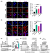
GABA(A) receptor activation enhances autophagic puncta and the flux and triggers multimerization of GABARAP and Nix. (A) Shown are confocal immunofluorescence microscopic images of H1792 cells under various treatments: DMSO or control; AM-101 (3 µM); radiation (RT); and AM-101 plus RT (combo) (scale bar, 20 µm). Radiation dose was 3 Gy. Cells were stained for DNA with DAPI (blue fluorescent) and LC3B (left) using LC3B antibody. LC3B puncta were quantified per 3 cells for each experimental group and plotted, as shown in the bar graph, where * p = 0.0108 (control vs. AM-101); ** p = 0.0037 (control vs. RT); **** p < 0.0001 (control vs. combo) (right), which reveals a similar effect between AM-101 versus RT, but combining these two has a statistically pronounced impact on puncta formation. (B) Confocal immunofluorescence microscopic images of H1792 cells under various treatments that were then stained for DAPI and Nix to identify and quantify Nix puncta (left). Nix puncta were quantified per 3 cells for each experimental group and plotted, as shown in the bar graph, where *** p = 0.0008 (control vs. AM-101); ** p = 0.006 (control vs. RT); **** p < 0.0001 (control vs. combo) (right), which reveals a pronounced effect of AM-101 on puncta formation and an increase in puncta when AM-101 is combined with radiation (RT) (combo treatment group). For statistical calculations, one-way ANOVA was performed and followed up with Dunnett’s multiple comparisons test. (C) Immunoblotting (using 4–15% gradient gel PAGE) demonstrates enhanced autophagic flux with LC3B-II as a marker in H1792 cells following co-treatment with AM-101 and bafilomycin A1. A representative immunoblot probed with an LC3B antibody shows the results from cell lysates of control (DMSO-treated) and three treatment groups: BafA1 alone, AM-101 alone, and AM-101 combined with BafA1. H1792 cells were treated with 3 μM AM-101 for 48 h, followed by either 50 nM bafilomycin A1 (AM-101 + BafA1) or DMSO (vehicle) for an additional 4 h. Control cells were treated with DMSO for 48 h and then with either 50 nM bafilomycin A1 or DMSO for 4 h. The right panel shows LC3B-II band intensities quantified using ImageJ, with bar graphs representing the fold increase in LC3B-II for each treatment group relative to the vehicle control (data shown as mean ± SEM, n = 2). Co-treatment of AM-101 and bafilomycin A1 significantly increased LC3B-II compared to AM-101 or bafilomycin A1 alone. GAPDH is used as loading control. To measure the statistical significance, ordinary one-way ANOVA (one-way ANOVA p < 0.0006) with Tukey’s multiple comparison test was performed to compare the means of each group. ** p = 0.0051 (BafA1 vs. AM-101); ** p = 0.0029 (BafA1 vs. AM-101 + BafA1); and *** p = 0.0005 (AM-101 vs. AM-101 + BafA1). (D) Modified immunoblotting of SDS gels (4–15% gradient gel) showing the effect of AM-101 on monomeric GABARAP expression and its oligomeric state in H1792 cells. (E) Modified immunoblotting of SDS gels (4–15% gradient gel) showing the effect of AM-101 on monomeric Nix protein expression and its oligomeric state in H1792 cells. AM-101 (3.0 µM) triggers a multimerization of GABARAP and an apparent increase in abundance at 72 h (left). Nix abundance is also enhanced as well as formation of dimer by AM-101 (3 µM) in a concentration-dependent manner in H1792 cells. GAPDH is used as a loading control for both experiments in (D,E). D: dimer; M: monomer. Original western blots are presented in Figure S9.
Figure 5
Figure 5
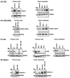
Change in abundance or utilization of autophagy biomarkers in response to GABA(A) activation. (A) ATG7 immunoblot (4–15% gradient polyacrylamide gel) of patient-derived lung adenocarcinoma primary (H1792) and brain-metastatic (UW-lung-16) cells following treatment with AM-101. Left, immunoblot showing increased expression of one isoform of ATG7 protein in primary H1792 cells of following treatment with 3.0 µM of AM-101 for 48 h compared to the control. Control: DMSO treated. Right, immunoblot showing change in ATG7 protein levels over time in lung cancer brain-metastatic UW-lung-16 cells treated with 3.0 µM AM-101 compared to the control. Significant increase in ATG7 protein is observed at 72 h. Control: DMSO treated. (B) Immunoblots showing the effect of in vitro AM-101 treatment on protein levels of ATG-12-ATG5 conjugate in primary H1792 cells (left panel) and patient-derived lung cancer brain metastatic UW-lung-16 (right panel) cells. In case of H1792 cells (left panel) sample was collected at 72 h after AM-101 treatment and in case of UW-lung-16 cells (right panel), samples were collected from both 48 h and 72 time points post treatment. (C) p62 immunoblots of SDS gels of lung adenocarcinoma primary (H1792) and brain-metastatic (UW-lung-16) cells following treatment with AM-101. Left, changes in expression of p62 protein as assessed by immunoblotting of lysates from H1792 cells treated with AM-101, radiation (3 Gy), and a combination of radiation (RT) plus AM-101 only. Control, DMSO. Middle, evaluation of the time-dependent utilization of p62 after AM-101 (3.0 µM) treatment in primary lung cancer cell (H1792). Right, patient-derived brain-metastatic lung adenocarcinoma cell line UW-lung-16 (right). Control, DMSO. GAPDH is used as a loading control. (D) Left, immunoblot showing Beclin-1 protein levels in control and AM-101 treated H1792 cells (treated for 72 h). Control, DMSO. Right, immunoblot shows Beclin-1 protein levels in patient-derived lung brain-metastatic UW-lung-16 cells treated with AM-101, radiation (3 Gy), and radiation (3 Gy) along with AM-101. Original western blots are presented in Figure S10.
Figure 6
Figure 6
GABARAP–Nix abrogation inhibits AM-101 cytotoxicity. (A) Combined treatment of lung adenocarcinoma (H1792) cells with AM-101 plus Pen3-ortho (P3o), a stapled-peptide that binds to GABARAP and abrogates Nix binding, inhibits the cytotoxicity of AM-101. The inhibitory effect of P3o is enhanced with an increased concentration of the inhibitor. **** p < 0.0001 [AM-101 vs. P3o (15 μM) + AM-101]; **** p < 0.0001 [AM-101 vs. P3o (25 μM) + AM-101]; **** p < 0.0001 [P3o (25 μM) vs. P3o (25 μM) + AM-101]. One-way ANOVA with Tukey’s multiple comparisons test was performed. (B) Treatment of H1792 cells with two different concentrations of P3o does not impact GABARAP protein abundance, as observed by immunoblot of SDS gel probed for GABARAP. (C) Treatment of H1792 cells with P3o reduces both Nix dimer and monomer protein levels, as observed by immunoblot of SDS gel probed for Nix. D: dimer, M: monomer. GAPDH is used as a loading control. Original western blots are presented in Figure S11.
Figure 7
Figure 7
Model of GABA(A)-receptor-mediated autophagy. (A) NSCLC cells possess intrinsic GABA(A) receptors (chloride anion channels). (B) AM-101 activates GABA(A) receptors, which in turn depolarize mitochondria. Changes in the cancer cell by binding of AM-101 to the receptor in combination with radiation include (i) enhanced expression–abundance of key genes involved in autophagy, including ATG7 and BECLIN-1; (ii) increased phosphorylation of the histone variant H2AX to generate γ-H2AX. (C) Depolarization induces key autophagic events in synergy with radiation: (i) enhanced expression and dimerization of GABA(A) receptor-associated protein, GABARAP; (ii) stabilization and dimerization of Nix, coupling GABARAP to mitochondria; (iii) enhanced expression of autophagy-associated proteins Beclin-1 and ATG7; (iv) utilization of ubiquitin-binding protein p62. Nix dimerization increases its stability and coordinates the nucleation of autophagosome formation. In this manner, GABA(A) receptor activation induces complex multimerization, activating autophagy. (D) Over time, GABARAP multimerizes commensurate with multimerization of the GABA(A) receptor, which enhances its activity. Created with BioRender.com.

Update of

References

    1. Siegel R.L., Miller K.D., Fuchs H.E., Jemal A. Cancer statistics, 2022. CA Cancer J. Clin. 2022;72:7–33. doi: 10.3322/caac.21708. - DOI - PubMed
    1. Fuchs J., Fruh M., Papachristofilou A., Bubendorf L., Hauptle P., Jost L., Zippelius A., Rothschild S.I. Resection of isolated brain metastases in non-small cell lung cancer (NSCLC) patients—Evaluation of outcome and prognostic factors: A retrospective multicenter study. PLoS ONE. 2021;16:e0253601. doi: 10.1371/journal.pone.0253601. - DOI - PMC - PubMed
    1. Sorensen J.B., Hansen H.H., Hansen M., Dombernowsky P. Brain metastases in adenocarcinoma of the lung: Frequency, risk groups, and prognosis. J. Clin. Oncol. 1988;6:1474–1480. doi: 10.1200/JCO.1988.6.9.1474. - DOI - PubMed
    1. Waqar S.N., Samson P.P., Robinson C.G., Bradley J., Devarakonda S., Du L., Govindan R., Gao F., Puri V., Morgensztern D. Non-small-cell lung cancer with brain metastasis at presentation. Clin. Lung Cancer. 2018;19:e373–e379. doi: 10.1016/j.cllc.2018.01.007. - DOI - PMC - PubMed
    1. Baschnagel A.M., Elnaggar J.H., VanBeek H.J., Kromke A.C., Skiba J.H., Kaushik S., Abel L., Clark P.A., Longhurst C.A., Nickel K.P., et al. ATR inhibitor M6620 (VX-970) enhances the effect of radiation in non-small cell lung cancer brain metastasis patient-derived xenografts. Mol. Cancer Ther. 2021;20:2129–2139. doi: 10.1158/1535-7163.MCT-21-0305. - DOI - PMC - PubMed

LinkOut - more resources