. 2024 Sep 17;16(18):3177.
doi: 10.3390/cancers16183177.

Protein Structure Inspired Discovery of a Novel Inducer of Anoikis in Human Melanoma

Affiliations

Protein Structure Inspired Discovery of a Novel Inducer of Anoikis in Human Melanoma

Fangfang Qiao et al. Cancers (Basel). .

Abstract

Drug discovery historically starts with an established function, either that of compounds or proteins. This can hamper discovery of novel therapeutics. As structure determines function, we hypothesized that unique 3D protein structures constitute primary data that can inform novel discovery. Using a computationally intensive physics-based analytical platform operating at supercomputing speeds, we probed a high-resolution protein X-ray crystallographic library developed by us. For each of the eight identified novel 3D structures, we analyzed binding of sixty million compounds. Top-ranking compounds were acquired and screened for efficacy against breast, prostate, colon, or lung cancer, and for toxicity on normal human bone marrow stem cells, both using eight-day colony formation assays. Effective and non-toxic compounds segregated to two pockets. One compound, Dxr2-017, exhibited selective anti-melanoma activity in the NCI-60 cell line screen. In eight-day assays, Dxr2-017 had an IC50 of 12 nM against melanoma cells, while concentrations over 2100-fold higher had minimal stem cell toxicity. Dxr2-017 induced anoikis, a unique form of programmed cell death in need of targeted therapeutics. Our findings demonstrate proof-of-concept that protein structures represent high-value primary data to support the discovery of novel acting therapeutics. This approach is widely applicable.

Keywords: anoikis; computational biology; drug discovery; protein structure.


Conflict of interest statement

The authors declare no conflicts of interest.

Figures

Figure 1
Figure 1
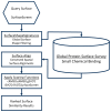
Computational pipeline schema for evaluating compound binding.
Figure 2
Figure 2
Schema for identification and characterization of 3D protein structures with the potential to bind therapeutically active small molecules.
Figure 3
Figure 3
Proteins that contain pockets structurally suited to binding drug-like small molecules. The surfaces of potential binding pockets are depicted, as are ribbon structures of proximal portions of the protein. The proteins are: 1-deoxy-D-xylulose 5-phosphate reductoisomerase (Dxr; Dxr1 (magenta) and Dxr2 (green) binding pockets), ß-ketoacyl acyl carrier protein reductase (FabG), 3-phosphoshikimate 1-carboxyvinyltransferase (EPSP synthase), dihydrofolate synthase (FolC; FolC1 (orange) and FolC2 (light green) binding pockets), hypoxanthine-guanine phosphoribosyltransferase (HGPRT), and glucose-1-phosphate thymidylyltransferase (TYLT).
Figure 4
Figure 4
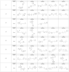
Structures of the acquired compounds.
Figure 5
Figure 5
Effect of 5FU on normal bone marrow. (A) Effect of 5FU on eight- and fourteen-day human stem cell hematopoietic colony formation. (B) Effect of 5FU on eight-day colony formation for human stem cells and HT29 colon cancer cells. Data are the mean ± SEM of N = 4 and N = 2 replicates for stem and HT29 cells, respectively.
Figure 6
Figure 6
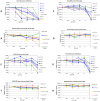
Effect of compounds on cancer cell and bone marrow colony formation. The effect of denoted compounds on eight-day cancer cell colony formation (A,C,E,G). The effect of denoted compounds on eight- and fourteen-day bone marrow colony formation (B,D,F,H). Data are mean ± SD (N = 2 replicates), with similar findings in separate experiments (also N = 2 replicates).
Figure 7
Figure 7
The predicted poses of bound Dxr2-017 and FolC2-001. The poses of FolC2-001 (A) and Dxr2-017 (B) bound to their respective FolC2 (yellow) and Dxr2 (green) binding surfaces are depicted, as are ribbon structures of proximal portions of the protein.
Figure 8
Figure 8
Dxr2-017 inhibits melanoma cell growth through induction of anoikis. (A) Inhibition of melanoma cell growth. Cells were treated with different concentrations of Dxr2-017, and formation of colonies at eight days is depicted. Data are expressed as the percent of untreated control and are the mean ± SEM of three separate experiments. Each experiment was conducted with N = 3 replicates. (B) No effect on cell cycle progression. M14 and SK-MEL-5 cells were treated for eight days with 20 nM and 50 nM of Dxr2-017, respectively, and the phase of the cell cycle was determined by flow cytometry. Control cells were treated with DMSO vehicle only. Representative histograms are depicted. Graphical data are the mean ± SEM (N = 3). * Denotes p-value ≤ 0.05. (C,D) Transition to floating cells. Cells were treated for three days with different Dxr2-017 concentrations. (C) Depicted are representative light photomicrographs at 20X. The scale bar is 150 µm. Blue and green arrows denote adherent and floating cells, respectively. (D) Adherent and floating cells were quantified from captured images. Data are the mean ± SEM (N = 4), expressed as a percentage to total cells. * Denotes p-value ≤ 0.05 compared to respective floating or attached cells. (E) Cleaved caspase 3 is induced in floating cells. Cells were treated for 3 days, and cell lysate from only adherent cells, or from adherent and floating cells combined, was probed by Western blot for cleaved caspase 3. (F) Dxr2-017 decreases cadherin. Cell lysate from only adherent cells or from adherent and floating cells combined was probed by Western blot for pan-cadherin. All experiments were repeated at separate times, yielding similar results. Original western blots are presented in File S2.

References

    1. Zhung W., Kim H., Kim W.Y. 3D molecular generative framework for interaction-guided drug design. Nat. Commun. 2024;15:2688. doi: 10.1038/s41467-024-47011-2. - DOI - PMC - PubMed
    1. Sakuma K., Kobayashi N., Sugiki T., Nagashima T., Fujiwara T., Suzuki K., Kobayashi N., Murata T., Kosugi T., Tatsumi-Koga R., et al. Design of complicated all-alpha protein structures. Nat. Struct. Mol. Biol. 2024;31:275–282. doi: 10.1038/s41594-023-01147-9. - DOI - PMC - PubMed
    1. Seoane B., Carbone A. The complexity of protein interactions unravelled from structural disorder. PLoS Comput. Biol. 2021;17:e1008546. doi: 10.1371/journal.pcbi.1008546. - DOI - PMC - PubMed
    1. Yao B., Li Z., Ding M., Chen M. Three-dimensional protein model similarity analysis based on salient shape index. BMC Bioinform. 2016;17:131. doi: 10.1186/s12859-016-0983-z. - DOI - PMC - PubMed
    1. Batool M., Ahmad B., Choi S. A Structure-Based Drug Discovery Paradigm. Int. J. Mol. Sci. 2019;20:2783. doi: 10.3390/ijms20112783. - DOI - PMC - PubMed

LinkOut - more resources