

Applying the Techniques of Materials Science towards an Understanding of the Process of Canine Intervertebral Disc Degeneration
- PMID: 39335255
- PMCID: PMC11428788
- DOI: 10.3390/ani14182665
Applying the Techniques of Materials Science towards an Understanding of the Process of Canine Intervertebral Disc Degeneration
Abstract
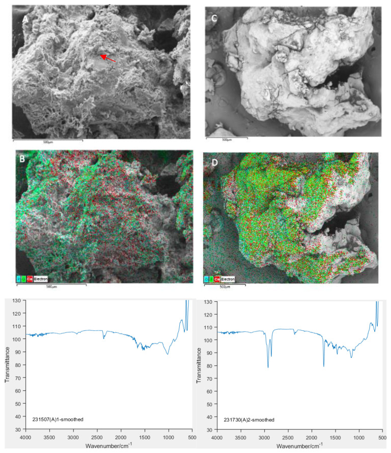
Intervertebral disc degeneration in dogs occurs in an accelerated way and involves calcification, which is associated with disc herniation or extrusion. The degenerative process is complex and involves the transformation of collagen fibres, loss of proteoglycans and notochord cells and a reduction in water content; however, how these processes are linked to future disc extrusion remains unknown. We have employed techniques including Fourier Transform Infra-red Spectroscopy (FTIR), Scanning Electron Microscopy (SEM), Transmission Electron Microscopy (TEM), Uniaxial Compression Loading and Atomic Force Microscopy (AFM) in an attempt to gain a greater understanding of the degenerative process and its consequences on the physical properties of the disc. FTIR verified by TEM demonstrated that calcium phosphate exists in an amorphous state within the disc and that the formation of crystalline particles of hydroxyapatite occurs prior to disc extrusion. AFM identified crystalline agglomerates consistent with hydroxyapatite as well as individual collagen fibres. SEM enabled the identification of regions rich in calcium, phosphorous and oxygen and allowed the visualization of the topographical landscape of the disc. Compression testing generated stress/strain curves which will facilitate investigation into disc stiffness. Ongoing work is aimed at identifying potential areas of intervention in the degenerative process as well as further characterizing the role of calcification in disc extrusion.
Keywords: Hansen; atomic force microscopy; calcification; disc disease; extrusion; intervertebral disk; spinal cord.
Conflict of interest statement
The authors declare no conflicts of interest.
Figures


References
-
- Brisson B.A., Moffatt S.L., Swayne S.L., Parent J.M. Recurrence of thoracolumbar intervertebral disk extrusion in chondrodystrophic dogs after surgical decompression with or without prophylactic fenestration: 265 cases (1995–1999) J. Am. Vet. Med. Assoc. 2004;224:1808–1814. doi: 10.2460/javma.2004.224.1808. - DOI - PubMed
Grants and funding
LinkOut - more resources
Full Text Sources
Miscellaneous
