. 2024 Sep 3;12(9):2013.
doi: 10.3390/biomedicines12092013.

ALCAT1-Mediated Pathological Cardiolipin Remodeling and PLSCR3-Mediated Cardiolipin Transferring Contribute to LPS-Induced Myocardial Injury

Affiliations

ALCAT1-Mediated Pathological Cardiolipin Remodeling and PLSCR3-Mediated Cardiolipin Transferring Contribute to LPS-Induced Myocardial Injury

Dong Han et al. Biomedicines. .

Abstract

Cardiolipin (CL), a critical phospholipid situated within the mitochondrial membrane, plays a significant role in modulating intramitochondrial processes, especially in the context of certain cardiac pathologies; however, the exact effects of alterations in cardiolipin on septic cardiomyopathy (SCM) are still debated and the underlying mechanisms remain incompletely understood. This study highlights a notable increase in the expressions of ALCAT1 and PLSCR3 during the advanced stage of lipopolysaccharide (LPS)-induced SCM. This up-regulation potential contribution to mitochondrial dysfunction and cellular apoptosis-as indicated by the augmented oxidative stress and cytochrome c (Cytc) release-coupled with reduced mitophagy, decreased levels of the antiapoptotic protein B-cell lymphoma-2 (Bcl-2) and lowered cell viability. Additionally, the timing of LPS-induced apoptosis coincides with the decline in both autophagy and mitophagy at the late stages, implying that these processes may serve as protective factors against LPS-induced SCM in HL-1 cells. Together, these findings reveal the mechanism of LPS-induced CL changes in the center of SCM, with a particular emphasis on the importance of pathological remodeling and translocation of CL to mitochondrial function and apoptosis. Additionally, it highlights the protective effect of mitophagy in the early stage of SCM. This study complements previous research on the mechanism of CL changes in mediating SCM. These findings enhance our understanding of the role of CL in cardiac pathology and provide a new direction for future research.

Keywords: cardiolipin; pathological CL remodeling; septic cardiomyopathy; translocation of CL.


Conflict of interest statement

The authors declare no conflict of interest.

Figures
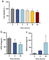
Figure 1
Figure 1
Changes in cell death and oxidative stress in the LPS-induced HL-1 cardiac cells: (a) cell viability was scored by methyl thiazolyl tetrazolium (MTT) assay, n = 5 independent experiments; (b) the level of SOD activity decreased in LPS-induced HL-1 cells, n = 3 independent experiments; (c) the level of MDA activity increased in LPS-induced HL-1 cells, n = 3 independent experiments. * p < 0.05, ** p < 0.01, compared with the control group.
Figure 2
Figure 2
Changes in autophagy and apoptosis in the LPS-induced HL-1 cardiac cells: (a) Western blot images showing the time-course change in LC3-II in LPS-induced HL-1 cells, n = 5 independent experiments; (b) Western blot images showing the time-course change in P62 in LPS-induced HL-1 cells, n = 4 independent experiments; (c) Western blot images showing the change in BCL2 in LPS-induced HL-1 cells, n = 4 independent experiments; (d) Western blot images showing the change in cytochrome c in LPS-induced HL-1 cells, n = 3 independent experiments; (e) LC3 aggregation quantified under confocal fluorescence microscopy. * p < 0.05, ** p < 0.01, compared with the control group.
Figure 3
Figure 3
Changes in mitophagy in the LPS-induced HL-1 cardiac cells. Representative TEM images of HL-1 cells in control, LPS-treated for 4 h, LPS-treated for 24 h. The images below showcase an enlarged view delineated by the dashed boundary. Arrows, formation of autophagosomes.
Figure 4
Figure 4
Changes in ALCAT1 protein and PLSCR3 protein in LPS-induced HL-1 cells: (a) Western blot images showing the time-course change in ALCAT1 in LPS-induced HL-1 cells, n = 4 independent experiments; (b) Western blot images showing the time-course change in PLSCR3 in LPS-induced HL-1 cells, n = 4 independent experiments. * p < 0.05, ** p < 0.01, compared with the control group.

References

    1. Singer M., Deutschman C.S., Seymour C.W., Shankar-Hari M., Annane D., Bauer M., Bellomo R., Bernard G.R., Chiche J.-D., Coopersmith C.M., et al. The Third International Consensus Definitions for Sepsis and Septic Shock (Sepsis-3) J. Am. Med. Assoc. 2016;315:801–810. doi: 10.1001/jama.2016.0287. - DOI - PMC - PubMed
    1. Hollenberg S.M., Singer M. Pathophysiology of sepsis-induced cardiomyopathy. Nat. Rev. Cardiol. 2021;18:424–434. doi: 10.1038/s41569-020-00492-2. - DOI - PubMed
    1. Parker M.M., Shelhamer J.H., Bacharach S.L., Green M.V., Natanson C., Frederick T.M., Damske B.A., Parrillo J.E. Profound but reversible myocardial depression in patients with septic shock. Ann. Intern. Med. 1984;100:483–490. doi: 10.7326/0003-4819-100-4-483. - DOI - PubMed
    1. Stanzani G., Duchen M.R., Singer M. The role of mitochondria in sepsis-induced cardiomyopathy. Biochim. Biophys. Acta Mol. Basis. Dis. 2019;1865:759–773. doi: 10.1016/j.bbadis.2018.10.011. - DOI - PubMed
    1. Chen T., Ye L., Zhu J., Tan B., Yi Q., Sun Y., Xie Q., Xiang H., Wang R., Tian J., et al. Inhibition of pyruvate dehydrogenase kinase 4 attenuates myocardial and mitochondrial injury in sepsis-induced cardiomyopathy. J. Infect. Dis. 2023;229:1178–1188. doi: 10.1093/infdis/jiad365. - DOI - PubMed

LinkOut - more resources