. 2024 Sep 7;12(9):2038.
doi: 10.3390/biomedicines12092038.

Investigation of High Frequency Irreversible Electroporation for Canine Spontaneous Primary Lung Tumor Ablation

Affiliations

Investigation of High Frequency Irreversible Electroporation for Canine Spontaneous Primary Lung Tumor Ablation

Alayna N Hay et al. Biomedicines. .

Abstract

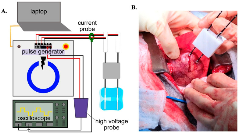
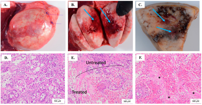
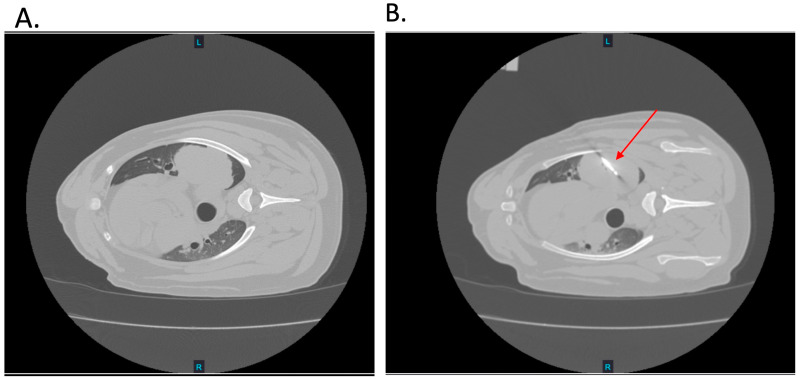
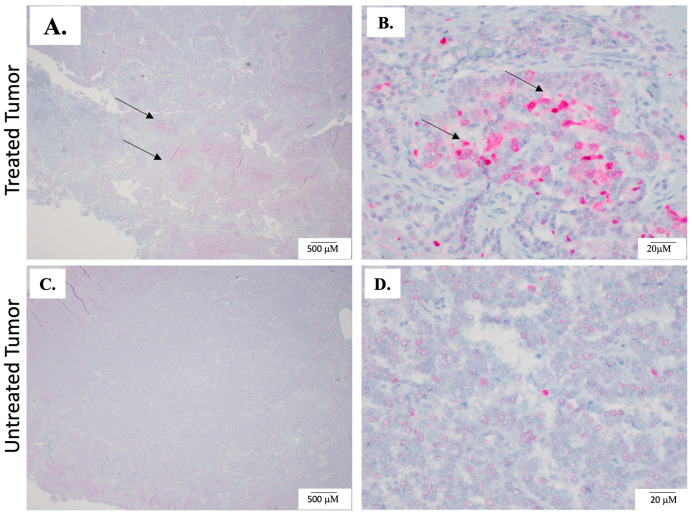
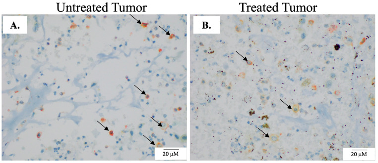
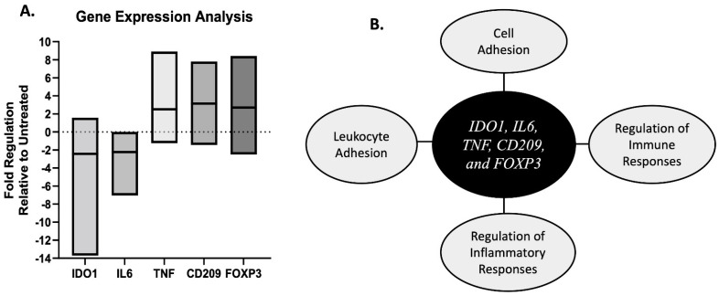
In this study, the feasibility of treating canine primary lung tumors with high-frequency irreversible electroporation (H-FIRE) was investigated as a novel lung cancer treatment option. H-FIRE is a minimally invasive tissue ablation modality that delivers bipolar pulsed electric fields to targeted cells, generating nanopores in cell membranes and rendering targeted cells nonviable. In the current study, canine patients (n = 5) with primary lung tumors underwent H-FIRE treatment with an applied voltage of 2250 V using a 2-5-2 µs H-FIRE waveform to achieve partial tumor ablation prior to the surgical resection of the primary tumor. Surgically resected tumor samples were evaluated histologically for tumor ablation, and with immunohistochemical (IHC) staining to identify cell death (activated caspase-3) and macrophages (IBA-1, CD206, and iNOS). Changes in immunity and inflammatory gene signatures were also evaluated in tumor samples. H-FIRE ablation was evident by the microscopic observation of discrete foci of acute hemorrhage and necrosis, and in a subset of tumors (n = 2), we observed a greater intensity of cleaved caspase-3 staining in tumor cells within treated tumor regions compared to adjacent untreated tumor tissue. At the study evaluation timepoint of 2 h post H-FIRE, we observed differential gene expression changes in the genes IDO1, IL6, TNF, CD209, and FOXP3 in treated tumor regions relative to paired untreated tumor regions. Additionally, we preliminarily evaluated the technical feasibility of delivering H-FIRE percutaneously under CT guidance to canine lung tumor patients (n = 2). Overall, H-FIRE treatment was well tolerated with no adverse clinical events, and our results suggest H-FIRE potentially altered the tumor immune microenvironment.

Keywords: canine oncology; comparative oncology; lung cancer; tumor ablation.


Conflict of interest statement

The authors declare the following conflict of interest: The author R.V.D. has ownership interest in the startup companies within the field of bioelectrics. R.V.D. also receives royalty income from technologies he has invented and serves as a consultant. Authors Lorenzo, Aycock, and Davalos have patents in the area of electroporation.

Figures

Figure 1
Figure 1
H-FIRE treatment set up and delivery. (A) Schematic of H-FIRE equipment. (B) Picture depicting the insertion of the bipolar electrodes into a canine lung tumor.
Figure 2
Figure 2
H-FIRE ablation histological outcomes. For all patients untreated and treated tumor samples were evaluated grossly and microscopically. (A) Sutures placed at the treated region to ensure positive identification of the ablation site. (B) The blue arrows indicate the treated regions of the tumor and (C) the blue arrows represent the treated region in a formalin fixed treated sample. (D) Representative untreated tumor region. (E,F) Representative images depicting variable but distinguished amounts of acute hemorrhage, which are the small red circles depicted in the treated region image (E) and throughout image (F). Prominent acute hemorrhage is not observed in image (D) or the untreated region in image (E). (F) Areas of tumor cell loss and replacement by fibrin are indicated by asterisks.
Figure 3
Figure 3
Percutaneous H-FIRE Delivery. (A) Representative CT image of patient lung tumor prior to H-FIRE probe insertion. (B) Representative CT image depicting the inserted H-FIRE probe into the tumor (red arrow).
Figure 4
Figure 4
Caspase-3 staining in tumor samples. To investigate the effects of H-FIRE on cell death, paired sections of untreated and treated samples of the tumor were evaluated for the apoptosis marker cleaved caspase-3 with IHC. (A,B) The dark pink/red staining denoted by arrows is positive cleaved caspase-3 staining and was most prominent in the cytoplasm of treated tumor cells (B) In general, treated tumor sections exhibited increased amounts of caspase-3 staining when compared to untreated areas. (C,D) Representative images of untreated tumor sections, which have minimal cleaved caspase-3 staining.
Figure 5
Figure 5
Macrophage infiltration post H-FIRE ablation. To evaluate the phenotype of infiltrating immune cells in paired untreated and treated sections of the tumor, multiplex IHC was performed. Our macrophage panel included IBA-1, CD206, and iNOS. Representative images of paired untreated (A) and H-FIRE-treated tumor regions (B). The co-staining of IBA-1 and CD206 is depicted by the orange color and arrows.
Figure 6
Figure 6
Differential gene expression analysis in paired treated and untreated tumor samples. (A) Box and whisker plot of fold regulation values in treated tumor samples relative to untreated tumor samples with a group average fold change of ≥2-fold or ≤−2-fold. (B) Diagram of gene ontology biological pathways associated with the five genes with a fold regulation value of ≥2-fold or ≤−2-fold.
Figure 7
Figure 7
In vitro analysis of H-FIRE ablation lesion size and lethal threshold. (A) Ablation lesion area 24 h post delivery of H-FIRE treatment to CLAC hydrogels in vitro. (B) Lethal electric field at each burst delivery rate parameter. * p < 0.05.

References

    1. Hahn F.F., Muggenburg B.A., Griffith W.C. Primary lung neoplasia in a beagle colony. Vet. Pathol. 1996;33:633–638. doi: 10.1177/030098589603300601. - DOI - PubMed
    1. Dobson J.M., Samuel S., Milstein H., Rogers K., Wood J.L. Canine neoplasia in the UK: Estimates of incidence rates from a population of insured dogs. J. Small Anim. Pract. 2002;43:240–246. doi: 10.1111/j.1748-5827.2002.tb00066.x. - DOI - PubMed
    1. de Groot P.M., Wu C.C., Carter B.W., Munden R.F. The epidemiology of lung cancer. Transl. Lung Cancer Res. 2018;7:220–233. doi: 10.21037/tlcr.2018.05.06. - DOI - PMC - PubMed
    1. Siegel R.L., Miller K.D., Jemal A. Cancer statistics, 2020. CA Cancer J. Clin. 2020;70:7–30. doi: 10.3322/caac.21590. - DOI - PubMed
    1. McPhetridge J.B., Scharf V.F., Regier P.J., Toth D., Lorange M., Tremolada G., Dornbusch J.A., Selmic L.E., Bae S., Townsend K.L., et al. Distribution of histopathologic types of primary pulmonary neoplasia in dogs and outcome of affected dogs: 340 cases (2010–2019) J. Am. Vet. Med. Assoc. 2021;260:234–243. doi: 10.2460/javma.20.12.0698. - DOI - PubMed

LinkOut - more resources