. 2024 Sep 18;12(9):2122.
doi: 10.3390/biomedicines12092122.

Exploring the Antibacterial and Regenerative Properties of a Two-Stage Alginate Wound Dressing in a Rat Model of Purulent Wounds

Affiliations

Exploring the Antibacterial and Regenerative Properties of a Two-Stage Alginate Wound Dressing in a Rat Model of Purulent Wounds

Aliya Atepileva et al. Biomedicines. .

Abstract

Chronic wounds complicated by infection pose significant clinical challenges, necessitating comprehensive treatment approaches. The widespread use of antibiotics has led to resistant microorganisms, complicating traditional therapies. This study aims to develop and evaluate modified alginate wound dressings with enhanced antimicrobial and regenerative properties. Alginate dressings were synthesized with silver nanoparticles, cefepime, and fibroblast growth factor-2 (FGF-2). The two-stage therapy involved an initial antibacterial dressing followed by a regenerative dressing. In vitro tests demonstrated high antibacterial activity, with maximum inhibition zones for P. aeruginosa (41.3 ± 0.4 mm) and S. aureus (36.6 ± 1.8 mm). In vivo studies on rats with purulent wounds showed significant healing progression in the experimental group. Histological analysis revealed complete re-epithelialization, thicker neoepithelium, dense collagen deposition, and minimal inflammation in treated wounds. These findings suggest that the modified alginate dressings significantly enhance the reparative process and are promising for treating chronic infected wounds in both veterinary and medical practices.

Keywords: FGF-2; alginate wound dressings; antibacterial properties; cefepime; chronic wounds; regenerative medicine; silver nanoparticles.


Conflict of interest statement

The authors declare no conflicts of interest.

Figures
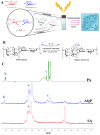
Figure 1
Figure 1
Synthesis and characterization of alginate hydrogel. (A) General graphical illustration of the synthesis process for the modified alginate hydrogel. (B) Schematic illustration of the sodium alginate hydrogel modification. Please note that the schematic structure shows only one possible pathway of the click chemistry reaction. In reality, the reaction could occur with any hydroxyl groups present in the sodium alginate. (C) 1H NMR spectra of sodium alginate (Alg), 4—pentenoic anhydride (PA), and their resulting product (AlgP) recorded in D2O at 50 °C. (D) Illustration of the alginate hydrogel disk. (E) SEM image of the surface morphology of the alginate hydrogel. Scale bar: 30 µm.
Figure 1
Figure 1
Synthesis and characterization of alginate hydrogel. (A) General graphical illustration of the synthesis process for the modified alginate hydrogel. (B) Schematic illustration of the sodium alginate hydrogel modification. Please note that the schematic structure shows only one possible pathway of the click chemistry reaction. In reality, the reaction could occur with any hydroxyl groups present in the sodium alginate. (C) 1H NMR spectra of sodium alginate (Alg), 4—pentenoic anhydride (PA), and their resulting product (AlgP) recorded in D2O at 50 °C. (D) Illustration of the alginate hydrogel disk. (E) SEM image of the surface morphology of the alginate hydrogel. Scale bar: 30 µm.
Figure 2
Figure 2
Evaluation of FGF-2 release and cytotoxicity of alginate hydrogels. (A) Profile of cumulative FGF-2 release from Alg-Ca-FGF-2 and Alg-FGF-2 hydrogels. (B) MTT assay for cytotoxicity of alginate hydrogels on rat ADMSC cells. (C) The effect of dressings with FGF-2, cefepime, and silver nanoparticles on the growth of dermal fibroblasts. Positive controls: FGF-2 (100 ng/mL), cefepime (10 mg/mL), and AgNPs (10 mg/mL) in culture medium. Values are presented as the mean value ± SD (n = 5). * p < 0.05, ** p < 0.01.
Figure 3
Figure 3
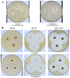
Antibacterial activity of alginate hydrogels. (A) Titer of cells from the overnight cultures of S. aureus and P. aeruginosa determined by visual counting of colonies after inoculation on LB culture medium. (B) Inhibition zones of test cultures of S. aureus and P. aeruginosa after incubation with hydrogel samples containing silver particles and cefepime at a concentration of 1%. The values are presented as the mean value ± SD (n = 5).
Figure 4
Figure 4
Regeneration of epidermal tissue in purulent wounds of rats using two–stage alginate dressing therapy. (A) Microscopic images of purulent wounds at 3, 7, and 14 days. (B) Wound healing rates in control and experimental groups. (C) Results of bacterial cultures taken from wounds on days 1, 3, and 7. * p < 0.05, ** p < 0.01. (D) Histological image of a wound area section: ED—epidermis; GT—granulation tissue; AT—adipose tissue; SA—skin appendage; robust, wellrevascularized GT (↓); poorly developed GT (↔); well—formed, dense ED (*); thin, fragile ED (**); regenerated AT (●).
Figure 4
Figure 4
Regeneration of epidermal tissue in purulent wounds of rats using two–stage alginate dressing therapy. (A) Microscopic images of purulent wounds at 3, 7, and 14 days. (B) Wound healing rates in control and experimental groups. (C) Results of bacterial cultures taken from wounds on days 1, 3, and 7. * p < 0.05, ** p < 0.01. (D) Histological image of a wound area section: ED—epidermis; GT—granulation tissue; AT—adipose tissue; SA—skin appendage; robust, wellrevascularized GT (↓); poorly developed GT (↔); well—formed, dense ED (*); thin, fragile ED (**); regenerated AT (●).

References

    1. Shmatenko O.P., Pidlisny O.V., Prykhodko T.V., Solomennyi A.M., Pritula R.L., Semenchenko G.B., Takhtaulova N.O. Technological aspects of creating soft dosage forms for the treatment of purulent wounds. Ukr. J. Mil. Med. 2020;1:50–63. doi: 10.46847/ujmm.2020.1(1)-050. - DOI
    1. Zubarev P.N., Ivanus S.Y., Risman B. V Modern Principles of Treatment of Purulent Wounds. Volume 37 Food and Agriculture Organization of the United Nations; Rome, Italy: 2017.
    1. Guo S., Dipietro L.A. Factors Affecting Wound Healing. J. Dent. Res. 2010;89:219–229. doi: 10.1177/0022034509359125. - DOI - PMC - PubMed
    1. Medzhitov R. Origin and Physiological Roles of Inflammation. Nature. 2008;454:428–435. doi: 10.1038/nature07201. - DOI - PubMed
    1. Hotchkiss R.S., Karl I.E. The Pathophysiology and Treatment of Sepsis. N. Engl. J. Med. 2003;348:138–150. doi: 10.1056/NEJMra021333. - DOI - PubMed

LinkOut - more resources