

Review of In Situ Hybridization (ISH) Stain Images Using Computational Techniques
- PMID: 39335767
- PMCID: PMC11430898
- DOI: 10.3390/diagnostics14182089
Review of In Situ Hybridization (ISH) Stain Images Using Computational Techniques
Abstract
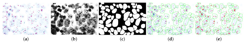
Recent advancements in medical imaging have greatly enhanced the application of computational techniques in digital pathology, particularly for the classification of breast cancer using in situ hybridization (ISH) imaging. HER2 amplification, a key prognostic marker in 20-25% of breast cancers, can be assessed through alterations in gene copy number or protein expression. However, challenges persist due to the heterogeneity of nuclear regions and complexities in cancer biomarker detection. This review examines semi-automated and fully automated computational methods for analyzing ISH images with a focus on HER2 gene amplification. Literature from 1997 to 2023 is analyzed, emphasizing silver-enhanced in situ hybridization (SISH) and its integration with image processing and machine learning techniques. Both conventional machine learning approaches and recent advances in deep learning are compared. The review reveals that automated ISH analysis in combination with bright-field microscopy provides a cost-effective and scalable solution for routine pathology. The integration of deep learning techniques shows promise in improving accuracy over conventional methods, although there are limitations related to data variability and computational demands. Automated ISH analysis can reduce manual labor and increase diagnostic accuracy. Future research should focus on refining these computational methods, particularly in handling the complex nature of HER2 status evaluation, and integrate best practices to further enhance clinical adoption of these techniques.
Keywords: deep learning; fluorescent in situ hybridization (FISH); human epidermal growth factor receptor 2 (HER2); pathologies; silver-enhanced in situ hybridization (SISH).
Conflict of interest statement
The authors declare no conflicts of interest.
Figures
References
-
- Coulton G.R., De Belleroche J. In Situ Hybridization: Medical Applications. Springer Science & Business Media; Berlin/Heidelberg, Germany: 2012.
-
- Di Palma S., Collins N., Faulkes C., Ping B., Ferns G., Haagsma B., Layer G., Kissin M., Cook M. Chromogenic in situ hybridisation (CISH) should be an accepted method in the routine diagnostic evaluation of HER2 status in breast cancer. J. Clin. Pathol. 2007;60:1067–1068. doi: 10.1136/jcp.2006.043356. - DOI - PMC - PubMed
-
- Shousha S., Peston D., Amo-Takyi B., Morgan M., Jasani B. Evaluation of automated silver-enhanced in situ hybridization (SISH) for detection of HER2 gene amplification in breast carcinoma excision and core biopsy specimens. Histopathology. 2009;54:248–253. doi: 10.1111/j.1365-2559.2008.03185.x. - DOI - PubMed
Publication types
LinkOut - more resources
Full Text Sources
Research Materials
Miscellaneous
