. 2024 Sep 18;13(18):2944.
doi: 10.3390/foods13182944.

Cardiac Hypertrophy in Pregnant Rats, Descendants of Fructose-Fed Mothers, an Effect That Worsens with Fructose Supplementation

Affiliations

Cardiac Hypertrophy in Pregnant Rats, Descendants of Fructose-Fed Mothers, an Effect That Worsens with Fructose Supplementation

Cristina Donis et al. Foods. .

Abstract

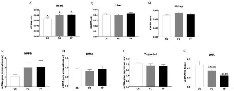
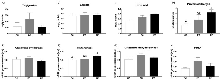
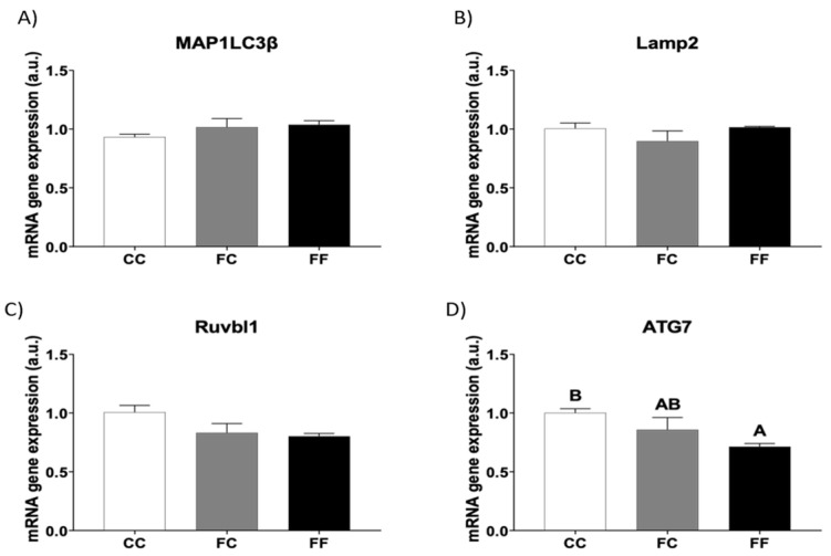
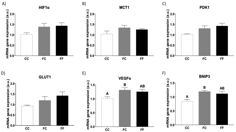
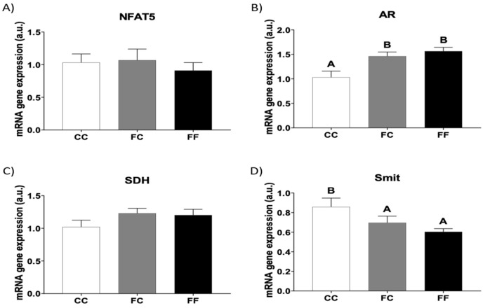
The role of fructose consumption in the development of obesity, MetS, and CVD epidemic has been widely documented. Notably, among other effects, fructose consumption has been demonstrated to induce cardiac hypertrophy. Moreover, fructose intake during pregnancy can cause hypertrophy of the maternal heart. Our previous research has demonstrated that maternal fructose intake has detrimental effects on fetuses, which persist into adulthood and are exacerbated upon re-exposure to fructose. Additionally, we found that maternal fructose consumption produces changes in female progeny that alter their own pregnancy. Despite these findings, fructose intake during pregnancy is not currently discouraged. Given that cardiac hypertrophy is a prognostic marker for heart disease and heart failure, this study aimed to determine whether metabolic changes occurring during pregnancy in the female progeny of fructose-fed mothers could provoke a hypertrophic heart. To test this hypothesis, pregnant rats from fructose-fed mothers, with (FF) and without (FC) fructose supplementation, were studied and compared to pregnant control rats (CC). Maternal hearts were analyzed. Although both FF and FC mothers exhibited heart hypertrophy compared to CC rats, cardiac DNA content was more diminished in the hearts of FF dams than in those of FC rats, suggesting a lower number of heart cells. Accordingly, changes associated with cardiac hypertrophy, such as HIF1α activation and hyperosmolality, were observed in both the FC and FF dams. However, FF dams also exhibited higher oxidative stress, lower autophagy, and decreased glutamine protection against hypertrophy than CC dams. In conclusion, maternal fructose intake induces changes in female progeny that alter their own pregnancy, leading to cardiac hypertrophy, which is further exacerbated by subsequent fructose intake.

Keywords: cardiac hypertrophy; fetal programming; fructose; pregnancy.


Conflict of interest statement

The authors declare no conflicts of interest.

Figures

Figure 1
Figure 1
Pregnancy in progeny from fructose-fed mothers and ingestion of liquid fructose throughout gestation in pregnant rats from fructose-fed mothers induce cardiac hypertrophy. (A) Heart weight corrected by BW, (B) liver weight corrected by BW, and (C) kidney weight corrected by BW of pregnant rats from control mothers (CC, empty bar) or pregnant rats from fructose-fed mothers subjected (FF, black bar) or not (FC, gray bar) to fructose intake throughout own pregnancy. Heart levels of specific mRNA for hypertrophy markers: (D) NPPB, (E) βMhC, and (F) Troponin I gene expression. (G) DNA content of the tissue. The fold decrease is indicated in percentages in comparison to the CC group. Relative target gene mRNA levels were measured by Real-Time PCR as explained in Materials and Methods, normalized to Rps29 levels, and expressed in arbitrary units (a.u.). Data are expressed as the mean ± S.E., n = 5 rats. Values not sharing a common letter are significantly different (p < 0.05). NPPB: Natriuretic peptide B; βMhC: β Myosin heavy chain; BW: Body weight (conceptus free).
Figure 2
Figure 2
Metabolic and oxidative stress parameters related to cardiac hypertrophy in pregnant rats. Heart levels of (A) triglycerides, (B) lactate, (C) uric acid, and (D) protein carbonyls of pregnant rats from control mothers (CC, empty bar) or pregnant rats from fructose-fed mothers subjected (FF, black bar) or not (FC, gray bar) to fructose intake throughout own pregnancy. Heart levels of specific mRNA for (E) glutamine synthetase, (F) glutaminase, (G) glutamate dehydrogenase, and (H) PDK4 gene expression. Relative target gene mRNA levels were measured by Real-Time PCR as explained in Materials and Methods, normalized to Rps29 levels, and expressed in arbitrary units (a.u.). Data are expressed as mean ± S.E., n = 5 rats. Values not sharing a common letter are significantly different (p < 0.05). PDK4: Pyruvate dehydrogenase kinase 4.
Figure 3
Figure 3
Fructose consumption during pregnancy affects the expression of autophagy-related genes in the hearts of pregnant rats from fructose-fed mothers. Heart levels of specific mRNA for: (A) MAP1LC3β, (B) Lamp2, (C) Ruvbl1, and (D) ATG7 gene expression of pregnant rats from control mothers (CC, empty bar) or pregnant rats from fructose-fed mothers subjected (FF, black bar) or not (FC, gray bar) to fructose intake throughout own pregnancy. Relative target gene mRNA levels were measured by Real-Time PCR as explained in Materials and Methods, normalized to Rps29 levels, and expressed in arbitrary units (a.u.). Data are expressed as mean ± S.E., n = 5 rats. Values not sharing a common letter are significantly different (p < 0.05). MAP1LC3B: Microtubule-associated protein 1 light chain 3 beta; Lamp2: Lysosomal-associated membrane protein 2; Ruvbl1: RuvB-like AAA ATPase 1; ATG7: Autophagy related 7.
Figure 4
Figure 4
Pregnancy in progeny from fructose-fed mothers and ingestion of liquid fructose throughout gestation in pregnant rats from fructose-fed mothers stimulates cardiac gene expression of HIF1α target genes. Heart levels of specific mRNA for (A) HIF1α, (B) MCT1, (C) PDK1, (D) GLUT1, (E) VEGFα, and (F) BNIP3 gene expression of pregnant rats from control mothers (CC, empty bar) or pregnant rats from fructose-fed mothers subjected (FF, black bar) or not (FC, gray bar) to fructose intake throughout own pregnancy. Relative target gene mRNA levels were measured by Real-Time PCR as explained in Materials and Methods, normalized to Rps29 levels, and expressed in arbitrary units (a.u.). Data are expressed as mean ± S.E., n = 5 rats. Values not sharing a common letter are significantly different (p < 0.05). HIF1α: Hypoxia-inducible factor 1 subunit alpha; MCT1: Monocarboxylate transporter 1; PDK1: Pyruvate dehydrogenase kinase 1; GLUT1: Glucose transporter 1; VEGFA: Vascular endothelial growth factor A; BNIP3: BCL2 interacting protein 3.
Figure 5
Figure 5
Pregnancy in progeny from fructose-fed mothers and ingestion of liquid fructose throughout gestation in pregnant rats from fructose-fed mothers influence the expression of cardiac genes related to osmolality. Heart levels of specific mRNA for (A) NFAT5, (B) AR, (C) SDH, and (D) Smit gene expression in pregnant rats from control mothers (CC, empty bar) or pregnant rats from fructose-fed mothers subjected (FF, black bar) or not (FC, gray bar) to fructose intake throughout own pregnancy. Relative target gene mRNA levels were measured by Real-Time PCR as explained in Materials and Methods, normalized to Rps29 levels, and expressed in arbitrary units (a.u.). Data are expressed as means ± S.E., n = 5 rats. Values not sharing a common letter are significantly different (p < 0.05). NFAT5: Nuclear factor of activated T cells 5; AR: Aldose reductase; SDH: Sorbitol dehydrogenase; Smit: Sodium/Myoinositol cotransporter.

Similar articles

References

    1. Dong Y., Li W., Yin J. The intestinal-hepatic axis: A comprehensive review on fructose metabolism and its association with mortality and chronic metabolic diseases. Crit. Rev. Food Sci. Nutr. 2023:1–14. doi: 10.1080/10408398.2023.2253468. - DOI - PubMed
    1. Jensen T., Abdelmalek M.F., Sullivan S., Nadeau K.J., Green M., Roncal C., Nakagawa T., Kuwabara M., Sato Y., Kang D.H., et al. Fructose and Sugar: A Major Mediator of Nonalcoholic Fatty Liver Disease. J. Hepatol. 2018;68:1063–1075. doi: 10.1016/j.jhep.2018.01.019. - DOI - PMC - PubMed
    1. Jung S., Bae H., Song W., Jang C. Dietary Fructose and Fructose-Induced Pathologies. Annu. Rev. Nutr. 2022;42:45–66. doi: 10.1146/annurev-nutr-062220-025831. - DOI - PMC - PubMed
    1. Zhang Y., Meng Y., Chang S., Zhang R., Shi C. High fructose causes cardiac hypertrophy via mitochondrial signaling pathway. Am. J. Transl. Res. 2016;8:4869–4880. - PMC - PubMed
    1. Oldfield C.J., Duhamel T.A., Dhalla N.S. Mechanisms for the transition from physiological to pathological cardiac hypertrophy. Can. J. Physiol. Pharmacol. 2020;98:74–84. doi: 10.1139/cjpp-2019-0566. - DOI - PubMed

LinkOut - more resources