

Mechanisms of Senescence and Anti-Senescence Strategies in the Skin
- PMID: 39336075
- PMCID: PMC11428750
- DOI: 10.3390/biology13090647
Mechanisms of Senescence and Anti-Senescence Strategies in the Skin
Abstract
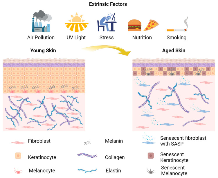
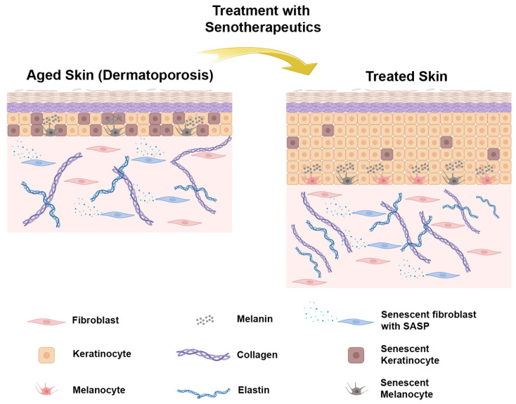
The skin is the layer of tissue that covers the largest part of the body in vertebrates, and its main function is to act as a protective barrier against external environmental factors, such as microorganisms, ultraviolet light and mechanical damage. Due to its important function, investigating the factors that lead to skin aging and age-related diseases, as well as understanding the biology of this process, is of high importance. Indeed, it has been reported that several external and internal stressors contribute to skin aging, similar to the aging of other tissues. Moreover, during aging, senescent cells accumulate in the skin and express senescence-associated factors, which act in a paracrine manner on neighboring healthy cells and tissues. In this review, we will present the factors that lead to skin aging and cellular senescence, as well as ways to study senescence in vitro and in vivo. We will further discuss the adverse effects of the accumulation of chronic senescent cells and therapeutic agents and tools to selectively target and eliminate them.
Keywords: SASP; cellular senescence; p16INK4a; senolytics; senomorphics; senotherapy; skin aging.
Conflict of interest statement
The authors declare no conflicts of interest.
Figures
Similar articles
-
Senolytics and senomorphics: Natural and synthetic therapeutics in the treatment of aging and chronic diseases.Free Radic Biol Med. 2021 Aug 1;171:169-190. doi: 10.1016/j.freeradbiomed.2021.05.003. Epub 2021 May 12. Free Radic Biol Med. 2021. PMID: 33989756 Review.
-
Skin senescence: mechanisms and impact on whole-body aging.Trends Mol Med. 2022 Feb;28(2):97-109. doi: 10.1016/j.molmed.2021.12.003. Epub 2022 Jan 7. Trends Mol Med. 2022. PMID: 35012887 Review.
-
Cellular Senescence in Diabetes Mellitus: Distinct Senotherapeutic Strategies for Adipose Tissue and Pancreatic β Cells.Front Endocrinol (Lausanne). 2022 Mar 31;13:869414. doi: 10.3389/fendo.2022.869414. eCollection 2022. Front Endocrinol (Lausanne). 2022. PMID: 35432205 Free PMC article. Review.
-
Exploring the fuzzy border between senolytics and senomorphics with chemoinformatics and systems pharmacology.Biogerontology. 2022 Aug;23(4):453-471. doi: 10.1007/s10522-022-09974-x. Epub 2022 Jul 4. Biogerontology. 2022. PMID: 35781578
-
Targeting Cellular Senescence with Senotherapeutics: Development of New Approaches for Skin Care.Plast Reconstr Surg. 2022 Oct 1;150:12S-19S. doi: 10.1097/PRS.0000000000009668. Epub 2021 Sep 28. Plast Reconstr Surg. 2022. PMID: 36170431 Free PMC article.
Cited by
-
Targeting Senescence: A Review of Senolytics and Senomorphics in Anti-Aging Interventions.Biomolecules. 2025 Jun 13;15(6):860. doi: 10.3390/biom15060860. Biomolecules. 2025. PMID: 40563501 Free PMC article. Review.
-
Comparison of the effects of fractional microneedle radiofrequency and microneedling on modulating the senescent fibroblast milieu in aged skin.Sci Rep. 2025 May 26;15(1):18296. doi: 10.1038/s41598-025-02545-3. Sci Rep. 2025. PMID: 40419589 Free PMC article. Clinical Trial.
-
Recent advances in dermal fibroblast senescence and skin aging: unraveling mechanisms and pioneering therapeutic strategies.Front Pharmacol. 2025 Jun 18;16:1592596. doi: 10.3389/fphar.2025.1592596. eCollection 2025. Front Pharmacol. 2025. PMID: 40606604 Free PMC article. Review.
-
From Bench to Bedside: Translating Cellular Rejuvenation Therapies into Clinical Applications.Cells. 2024 Dec 12;13(24):2052. doi: 10.3390/cells13242052. Cells. 2024. PMID: 39768144 Free PMC article. Review.
References
Publication types
Grants and funding
LinkOut - more resources
Full Text Sources
