Review
. 2024 Sep 3;13(9):690.
doi: 10.3390/biology13090690.

State of the Art in Sub-Phenotyping Midbrain Dopamine Neurons

Affiliations
Review

State of the Art in Sub-Phenotyping Midbrain Dopamine Neurons

Valentina Basso et al. Biology (Basel). .

Abstract

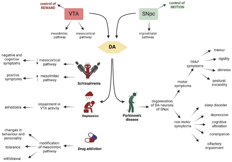
Dopaminergic neurons in the ventral tegmental area (VTA) and the substantia nigra pars compacta (SNpc) comprise around 75% of all dopaminergic neurons in the human brain. While both groups of dopaminergic neurons are in close proximity in the midbrain and partially overlap, development, function, and impairments in these two classes of neurons are highly diverse. The molecular and cellular mechanisms underlying these differences are not yet fully understood, but research over the past decade has highlighted the need to differentiate between these two classes of dopaminergic neurons during their development and in the mature brain. This differentiation is crucial not only for understanding fundamental circuitry formation in the brain but also for developing therapies targeted to specific dopaminergic neuron classes without affecting others. In this review, we summarize the state of the art in our understanding of the differences between the dopaminergic neurons of the VTA and the SNpc, such as anatomy, structure, morphology, output and input, electrophysiology, development, and disorders, and discuss the current technologies and methods available for studying these two classes of dopaminergic neurons, highlighting their advantages, limitations, and the necessary improvements required to achieve more-precise therapeutic interventions.

Keywords: Parkinson’s disease (PD); dopamine (DA); drug addiction; major depression; midbrain dopaminergic neurons (mDA); schizophrenia (SZ); substantia nigra pars compacta (SNpc); ventral tegmental area (VTA).


Conflict of interest statement

The authors declare no conflicts of interest.

Figures

Figure 1
Figure 1
Graphic representation of dopamine pathway in the brain. This pathway includes the mesolimbic pathway (pink) from the VTA to nucleus accumbens (orange, the mesocortical pathway (blue) from the VTA to cortex, and the nigrostriatal pathway (green) from SN to striatum. Created with BioRender (https://www.biorender.com/).
Figure 2
Figure 2
Graphic summary of the TFs involved in the development of mDA neurons that seem to have an impact in the differentiation of DA neurons of the VTA and SNpc. The expression of TFs plays roles in midbrain regional specification, specification and differentiation and maturation of the mDA phenotype, and it is exhibited across various embryonic (E) stages. The temporal line extends from E day 7 to beyond day 13. Arrows indicate a stimulatory effect, while perpendicular lines indicate an inhibitory effect of the TFs. TFs in black do not have a specific regional distribution pattern, while those written in red (VTA) or green (SNpc) colors indicate TFs whose regulation has a higher impact on the development or maintenance of VTA or SNpc DA neurons, respectively. Created with BioRender.
Figure 3
Figure 3
Schematic representation of the VTA on the left (in red), whose main role is the control of reward, and on the right (in green), of the SNpc, which controls motion. The VTA and the SNpc are linked to the production of DA (yellow). VTA is connected through the mesocortic pathway to the cortex and through the mesolimbic pathway to the NAc and striatum. Dysfunctions of these pathways are associated with schizophrenia, depression, and drug addiction. The SNpc controls the voluntary movement and projects via the nigrostriatal pathway to the dorsal striatum. Degeneration of DA neurons of the SNpc is associated with Parkinson’s disease. The characteristic symptoms of each pathology are indicated by arrows. Created with BioRender.
Figure 4
Figure 4
Schematic summary of all the differences between DA neurons of the VTA (in red) and the SNpc (in green). Created with BioRender.
Figure 5
Figure 5
Pros (in green) and cons (in red) of the samples used to investigate the characteristics of DA neurons and study the differences between the DA neurons of the VTA and SNpc. Created with BioRender.
Figure 6
Figure 6
Proteins found enriched in the midbrain and reported in the Allen Brain Atlas. The two highlighted genes are found in all five studies (Bossers et al., 2009 [232]; Yang et al., 2022 [233]; Verma et al., 2023 [234]; Zhou et al., 2023 [235]; and Huang et al., 2024 [236]).

References

    1. Montagu K.A. Catechol Compounds in Rat Tissues and in Brains of Different Animals. Nature. 1957;180:244–245. doi: 10.1038/180244a0. - DOI - PubMed
    1. Nagatsu T., Levitt M., Udenfriend S. Tyrosine Hydroxylase. The Initial Step in Norepinephrine Biosynthesis. J. Biol. Chem. 1964;239:2910–2917. doi: 10.1016/S0021-9258(18)93832-9. - DOI - PubMed
    1. Fernstrom J.D., Fernstrom M.H. Tyrosine, Phenylalanine, and Catecholamine Synthesis and Function in the Brain. J. Nutr. 2007;137:1539S–1547S. doi: 10.1093/jn/137.6.1539S. - DOI - PubMed
    1. Christenson J.G., Dairman W., Udenfriend S. Preparation and Properties of a Homogeneous Aromatic L-Amino Acid Decarboxylase from Hog Kidney. Arch. Biochem. Biophys. 1970;141:356–367. doi: 10.1016/0003-9861(70)90144-X. - DOI - PubMed
    1. Falck B., Hillarp N.A., Thieme G., Torp A. Fluorescence of Catechol Amines and Related Compounds Condensed with Formaldehyde. Brain Res. Bull. 1982;9:xi–xv. doi: 10.1016/0361-9230(82)90113-7. - DOI - PubMed

LinkOut - more resources