

Therapeutic Potential of Vitamin B Complex in Peripheral Nerve Injury Recovery: An Experimental Rat Model Study
- PMID: 39336597
- PMCID: PMC11434473
- DOI: 10.3390/medicina60091556
Therapeutic Potential of Vitamin B Complex in Peripheral Nerve Injury Recovery: An Experimental Rat Model Study
Abstract
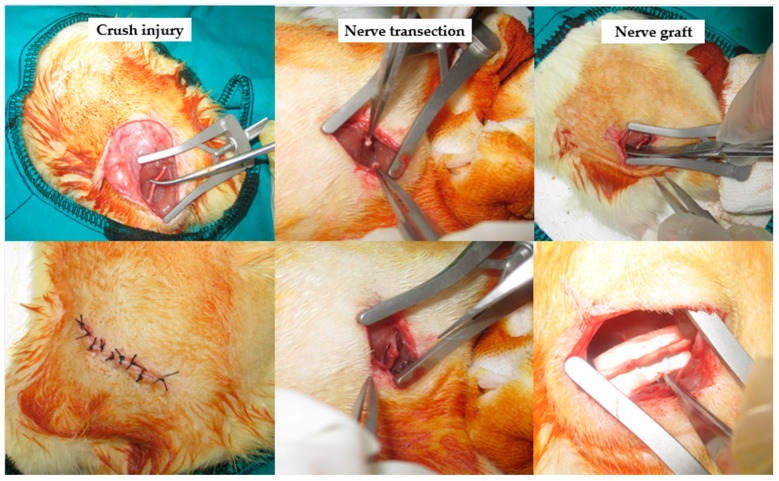
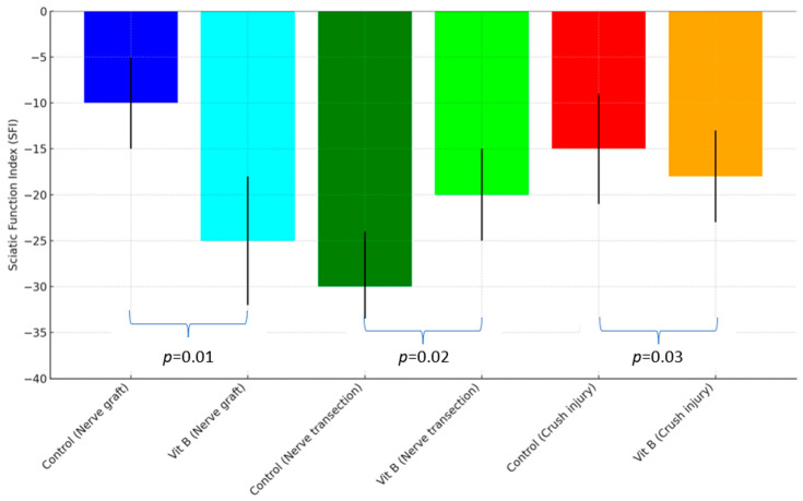
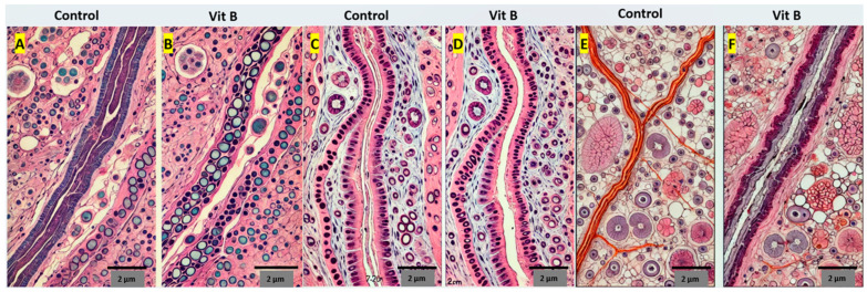
Objectives: Vitamin B complexes are frequently used in clinical practice for peripheral nerve trauma. However, there is a lack of scientific data on their effectiveness. This study aims to investigate the impact of the vitamin B complex on nerve recovery in a rat model of peripheral nerve paralysis. Materials and Methods: Sixty male Wistar Albino rats were divided into six groups. Models of nerve injury, including blunt trauma, nerve incision, and autograft, were performed on all rats approximately 1 cm distal to the sciatic notch. B-complex vitamins were injected intraperitoneally at 0.2 mL/day to the treatment groups. The control groups were given 0.2 mL/day saline. After 1 month, the study was terminated, electromyography (EMG) was performed to measure the conduction velocity, and nerve tissue was taken from the repair line. The sciatic function indexes (SFIs) were calculated and analyzed. The histopathological samples were stained with hematoxylin and eosin and Toluidine blue and examined with a light microscope. Pathologically, myelination, fibrosis, edema, and mast cell densities in the nervous tissue were evaluated. Results: The vitamin B treatment groups demonstrated significant improvements in SFI compared to the control groups, indicating functional improvement in nerve damage (p < 0.05). In the nerve graft group, the vitamin B group showed a shorter latency, higher velocity, and larger peak-to-peak compared to the controls (p < 0.05). In the nerve transection group, the vitamin B group had better latency, velocity, and peak-to-peak values than the controls (p < 0.05). In the crush injury group, the vitamin B group exhibited an improved latency, velocity, and peak-to-peak compared to the controls (p < 0.05). Better myelination, less fibrosis, edema, and mast cells were also in the vitamin B group (p < 0.05). Conclusions: Vitamin B treatment significantly improves nerve healing and function in peripheral nerve injuries. It enhances nerve conduction, reduces fibrosis, and promotes myelination, indicating its therapeutic potential in nerve regeneration.
Keywords: nerve healing; peripheral nerve injury; rat; vitamin B complex.
Conflict of interest statement
The authors declare no conflicts of interest.
Figures
References
-
- Mekaj A., Mekaj Y. The role of pharmacological agents in nerve regeneration after peripheral nerve repair. Peripher. Nerve Regen. Surg. New Ther. Approaches Incl. Biomater. Cell-Based Ther. Dev. 2017;10:151–174. doi: 10.5772/intechopen.68378. - DOI
-
- Naseri S., Samaram H., Naghavi N., Rassouli M.B., Mousavinezhad M. Types of Short-Duration Electrical Stimulation-Induced Efficiency in the Axonal Regeneration and Recovery: Comparative in Vivo Study in Rat Model of Repaired Sciatic Nerve and its Tibial Branch after Transection Injury. Neurochem. Res. 2024;49:2469–2479. doi: 10.1007/s11064-024-04154-4. - DOI - PubMed
MeSH terms
Substances
LinkOut - more resources
Full Text Sources
Medical
