

Anterior Segment Optical Coherence Tomography for the Tailored Treatment of Mooren's Ulcer: A Case Report
- PMID: 39336871
- PMCID: PMC11432291
- DOI: 10.3390/jcm13185384
Anterior Segment Optical Coherence Tomography for the Tailored Treatment of Mooren's Ulcer: A Case Report
Abstract
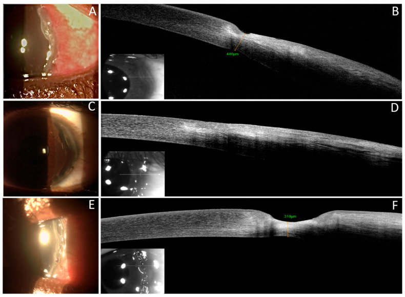
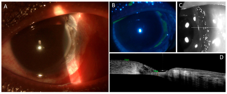
Background: Mooren's ulcer (MU) is a rare and debilitating form of peripheral ulcerative keratitis (PUK), characterized by a crescent-shaped ulcer with a distinctive overhanging edge at the corneal periphery. If left untreated, MU can lead to severe complications such as corneal perforation and blindness. Despite various treatment approaches, including anti-inflammatory and cytotoxic drugs, as well as surgical interventions, there is no clear evidence of the most effective treatment due to the lack of randomized controlled trials. AS-OCT is a non-invasive imaging technique that provides high-resolution cross-sectional images of the anterior segment, allowing for accurate evaluation of corneal ulcer characteristics, including depth, extent, and disease progression. Methods: We present the case of a 20-year-old male patient with MU managed using a stepladder approach, which included local and systemic corticosteroids, limbal conjunctival resection, and Cyclosporine A 1% eye drops. The patient underwent consecutive AS-OCT examinations and strict follow-up to tailor systemic and topical therapy. Results: Complete healing of the corneal ulcer with resolution of the inflammatory process was achieved. There was no recurrence of the disease at the 7-month follow-up. AS-OCT demonstrated progressive reorganization and thickening of the stromal tissue until the complete recovery of stromal thickness. Conclusions: The AS-OCT imaging modality allowed for the accurate evaluation of corneal ulcer characteristics, facilitating informed decision-making regarding the use of systemic immunosuppression, surgical interventions, and local immunomodulation and providing detailed and precise assessment of disease progression. This approach enabled a tailored and effective treatment strategy for the patient and played a critical role in guiding the therapeutic approach.
Keywords: AS-OCT; Mooren’s ulcer; PUK; corneal imaging; cyclosporine A.
Conflict of interest statement
The authors declare no conflicts of interest.
Figures

Similar articles
-
Bilateral Boston keratoprosthesis type 1 in a case of severe Mooren's ulcer.Eur J Ophthalmol. 2021 Mar;31(2):NP33-NP38. doi: 10.1177/1120672120909768. Epub 2020 Mar 6. Eur J Ophthalmol. 2021. PMID: 32141311
-
Evaluation of Mooren's corneal ulcer by anterior segment optical coherence tomography.Photodiagnosis Photodyn Ther. 2023 Dec;44:103806. doi: 10.1016/j.pdpdt.2023.103806. Epub 2023 Sep 16. Photodiagnosis Photodyn Ther. 2023. PMID: 37722614
-
A case of lattice corneal dystrophy type 1 with bilateral Mooren's ulcer.Am J Ophthalmol Case Rep. 2023 Jan 12;29:101796. doi: 10.1016/j.ajoc.2023.101796. eCollection 2023 Mar. Am J Ophthalmol Case Rep. 2023. PMID: 36718435 Free PMC article.
-
Rapid deterioration of Mooren's ulcers after conjunctival flap: a review of 2 cases.BMC Ophthalmol. 2017 Jun 15;17(1):93. doi: 10.1186/s12886-017-0488-1. BMC Ophthalmol. 2017. PMID: 28619029 Free PMC article. Review.
-
Treatment of Mooren's ulcer coexisting with a pterygium using an intrastromal lenticule obtained from small-incision lenticule extraction: case report and literature review.J Int Med Res. 2021 Jun;49(6):3000605211020246. doi: 10.1177/03000605211020246. J Int Med Res. 2021. PMID: 34130538 Free PMC article. Review.
Cited by
-
A Novel Pathogenic Variant in the KRT3 Gene in a Family with Meesmann Corneal Dystrophy.J Clin Med. 2025 Jan 28;14(3):851. doi: 10.3390/jcm14030851. J Clin Med. 2025. PMID: 39941522 Free PMC article.
References
-
- Tuft S. Mooren’s Ulcer. In: Johnson G.J., Minassian D.C., Weale R.A., West S.K., editors. Epidemiology of Eye Disease. Arnold; London, UK: 2003. pp. 209–211.
Publication types
LinkOut - more resources
Full Text Sources
Research Materials
