

Comparison of Dental Findings with Computed Tomographic and Clinical Examination in Patients with End-Stage Heart Failure
- PMID: 39336892
- PMCID: PMC11432535
- DOI: 10.3390/jcm13185406
Comparison of Dental Findings with Computed Tomographic and Clinical Examination in Patients with End-Stage Heart Failure
Abstract
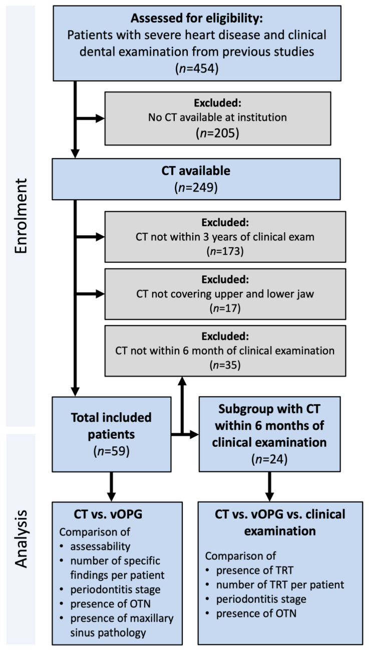
Background: This study aimed to evaluate the diagnostic value of pre-existing computed tomography (CT) examinations for the detection of dental pathologies compared with clinical dental examination in patients with end-stage heart failure. Methods: For this purpose, 59 patients with end-stage heart failure and pre-existing non-dental CT images of the craniofacial region were included. Virtual orthopantomograms (vOPG) were reconstructed. Dental pathologies were analyzed in vOPG and source-CT images. Imaging and clinical findings less than 6 months apart were compared (n = 24). Results: The subjective image quality of vOPG was more often rated as insufficient than CT (66%; 20%; p < 0.01). Depending on examination (CT, vOPG or clinic), between 33% and 92% of the patients could require dental intervention such as treatment of caries and periodontitis or tooth extraction. vOPG led to a higher (80%) prevalence of teeth requiring treatment than CT (39%; p < 0.01). The prevalence of teeth requiring treatment was similar in CT (29%) and clinic (29%; p = 1.00) but higher in vOPG (63%; p < 0.01). CT (stage 3 or 4: 42%) and vOPG (38%) underestimated the stage of periodontitis (clinic: 75%; p < 0.01). Conclusions: In conclusion, available CT images including the craniofacial region from patients with end-stage heart failure may contain valuable information regarding oral health status. The assessability of vOPGs might be insufficient and must be interpreted with caution.
Keywords: computed tomography; heart disease; heart transplantation; oral health; radiographs.
Conflict of interest statement
The authors declare no conflicts of interest.
Figures


References
-
- Bundesärztekammer (BÄK) Kassenärztliche Bundesvereinigung (KBV) National Disease Management Guideline: Chronic Heart Failure (in German) 2021. [(accessed on 29 May 2022)]. Available online: https://www.leitlinien.de/themen/herzinsuffizienz/3-auflage/kapitel-2.
-
- Seferović P.M., Vardas P., Jankowska E.A., Maggioni A.P., Timmis A., Milinković I., Polovina M., Gale C.P., Lund L.H., Lopatin Y., et al. The Heart Failure Association Atlas: Heart Failure Epidemiology and Management Statistics 2019. Eur. J. Heart Fail. 2021;23:906–914. doi: 10.1002/ejhf.2143. - DOI - PubMed
-
- Crespo-Leiro M.G., Metra M., Lund L.H., Milicic D., Costanzo M.R., Filippatos G., Gustafsson F., Tsui S., Barge-Caballero E., De Jonge N., et al. Advanced heart failure: A position statement of the Heart Failure Association of the European Society of Cardiology. Eur. J. Heart Fail. 2018;20:1505–1535. doi: 10.1002/ejhf.1236. - DOI - PubMed
Grants and funding
LinkOut - more resources
Full Text Sources
Medical
