Review
. 2024 Sep 19;13(18):5555.
doi: 10.3390/jcm13185555.

Silent Threats of the Heart: A Case Series and Narrative Review on Suicide Left Ventricle Post-Aortic Valve Replacement in Patients with Dynamic LVOT Obstruction and Aortic Stenosis

Affiliations
Review

Silent Threats of the Heart: A Case Series and Narrative Review on Suicide Left Ventricle Post-Aortic Valve Replacement in Patients with Dynamic LVOT Obstruction and Aortic Stenosis

Silvia Romano et al. J Clin Med. .

Abstract

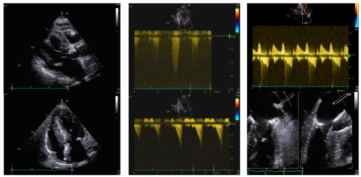
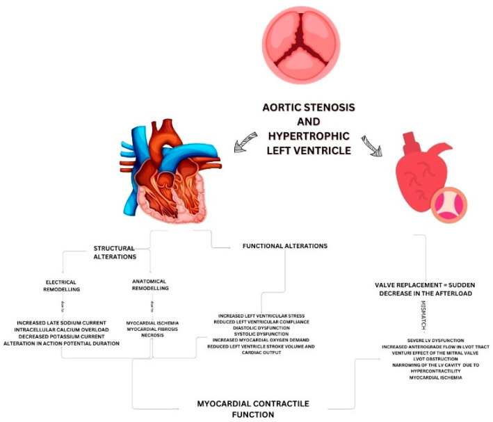
Aortic stenosis (AS) is the most prevalent valvular heart disease in Europe and North America, with transcatheter aortic valve implantation (TAVI) revolutionizing its management. Hypertrophic left ventricle (HLV) frequently coexists with AS, complicating treatment due to the associated risk of left ventricular outflow tract (LVOT) obstruction, heart failure, and sudden death. A rare but severe post-aortic valve replacement (AVR) complication, termed "suicide left ventricle" (SLV), has emerged, necessitating further study. This report synthesizes current literature on SLV, its pathophysiology, and management strategies, alongside four patient case studies. The patients aged 79-87 years, underwent AVR for symptomatic AS with HLV. Post-AVR, all experienced severe complications, including dynamicLVOT gradients, systolic anterior motion (SAM) of the mitral valve, and severe hypotension, leading to death in two cases. One patient survived following surgical aortic valve replacement (SAVR) with surgical myectomy. One patient survived after TAVI. These cases highlight the critical importance of multidisciplinary Heart Team evaluations and personalized treatment plans in managing SLV. Despite advancements in AVR, SLV remains a complex, life-threatening condition, requiring an exhaustive and multifaceted approach for optimal patient outcomes. This report offers valuable insights into SLV occurrence and management from a clinical perspective.

Keywords: aortic stenosis; hypertrophic left ventricle; multidisciplinary management; suicide left ventricle.


Conflict of interest statement

The authors declare no conflicts of interest.

Figures

Figure 1
Figure 1
Transthoracic and transesophageal echocardiography in an 87-year-old woman with paradoxical low-flow low-gradient aortic stenosis. Left panel. PLAX view (upper) and 4-chamber view (lower). IVS telediastolic diameter = 20 mm, LVEDVi = 31 mL/m2. Central panel. Aortic valve peak velocity of 3.6 m/s (upper) with a mean gradient of 28 mmHg and a dynamic LVOT gradient (lower) of 54 mmHg after the Valsalva maneuver. Right panel after TAVI. SAM of the anterior mitral leaflet (upper, red arrow) by TEE causing severe mitral regurgitation. IVS: interventricular septum. LVEDVi: left ventricular end-diastolic volume indexed. LVOT: left ventricular outflow tract. SAM: systolic anterior motion.
Figure 2
Figure 2
Transthoracic and transesophageal echocardiography in an 86-year-old woman before and after aortic valve stenosis percutaneous treatment and transcatheter alcohol septal ablation. Left panel: PLAX view (upper) and 4-chamber view (lower); IVS = 26 mm; LVEDVi = 45 mL/mq. Central panel: aortic valve peak velocity of 5.8 m/s (upper) with a mean gradient of 77 mmHg and dynamic LVOT gradient (lower) of 65 mmHg. Right panel—after TAVI and alcohol septal ablation: dynamic LVOT gradient (upper) of 20 mmHg but evidence of residual SAM of the anterior mitral leaflet (lower) by TEE. IVS: interventricular septum. LVEDVi: left ventricular end-diastolic volume indexed. LVOT: left ventricular outflow tract. SAM: systolic anterior motion. TAVI: transcatheter aortic valve implantation.
Figure 3
Figure 3
Transthoracic echocardiography of a 79-year-old woman before and after surgical aortic valve replacement and septal myectomy. Left panel. PLAX view (upper) and 4-chamber view (lower). IVS telediastolic diameter = 15 mm; LVEDVi 27 mL/m2. Central panel. Aortic valve peak velocity of 4.5 m/s (upper) with a mean gradient of 51 mmHg and dynamic LVOT gradient (lower) of 42 mmHg after the Valsalva maneuver. Right panel: after cardiac surgery. 4-chamber view (upper) and dynamic LVOT gradient of 11 mmHg (lower) after surgical aortic valve replacement. IVS: interventricular septum. LVEDVi: left ventricular end-diastolic volume indexed. LVOT: left ventricular outflow tract.
Figure 4
Figure 4
Transthoracic echocardiography of an 80-year-old man before and after aortic valve percutaneous replacement and pacemaker implantation. Left panel. PLAX view (upper) and 4-chamber view (lower). IVS telediastolic diameter = 18 mm; LVEDVi = 54 mL/m2. Central panel. Aortic valve peak velocity of 4.5 m/s (upper) with a mean gradient of 50 mmHg and dynamic LVOT gradient (lower) of 45 mmHg after the Valsalva maneuver. Right panel after TAVI. Increased dynamic LVOT gradient after TAVI (upper) of 65 mmHg after the Valsalva maneuver, which is reduced after pacemaker implantation (lower). IVS: interventricular septum. LVEDVi: left ventricular end-diastolic volume indexed. LVOT: left ventricular outflow tract. TAVI: transcatheter aortic valve implantation.
Figure 5
Figure 5
Pathophysiology of suicide left ventricle in patients with dynamic left ventricular outflow tract (LVOT) obstruction and aortic stenosis.
Figure 6
Figure 6
Red flags of suicide left ventricle and proposed strategy for hemodynamic optimization in patients with aortic stenosis and dynamic left ventricle outflow tract (LVOT) obstruction.

References

    1. Vahanian A., Beyersdorf F., Praz F., Milojevic M., Baldus S., Bauersachs J., Capodanno D., Conradi L., De Bonis M., De Paulis R., et al. 2021 ESC/EACTS Guidelines for the management of valvular heart disease. Eur. Heart J. 2022;43:561–632. doi: 10.1093/eurheartj/ehab395. - DOI - PubMed
    1. Vora A.N., Gada H., Manandhar P., Kosinski A., Kirtane A., Nazif T., Reardon M., Kodali S., Cohen D.J., Thourani V., et al. National Variability in Pacemaker Implantation Rate Following TAVR: Insights From the STS/ACC TVT Registry. JACC Cardiovasc. Interv. 2024;17:391–401. doi: 10.1016/j.jcin.2023.12.005. - DOI - PubMed
    1. Makkar R.R., Thourani V.H., Mack M.J., Kodali S.K., Kapadia S., Webb J.G., Yoon S.H., Trento A., Svensson L.G., Herrmann H.C., et al. Five-Year Outcomes of Transcatheter or Surgical Aortic-Valve Replacement. N. Engl. J. Med. 2020;382:799–809. doi: 10.1056/NEJMoa1910555. - DOI - PubMed
    1. Arbelo E., Protonotarios A., Gimeno J.R., Arbustini E., Barriales-Villa R., Basso C., Bezzina C.R., Biagini E., Blom N.A., de Boer R.A., et al. 2023 ESC Guidelines for the management of cardiomyopathies. Eur. Heart J. 2023;44:3503–3626. doi: 10.1093/eurheartj/ehad194. - DOI - PubMed
    1. Huded C.P., Arnold S.V., Cohen D.J., Manandhar P., Vemulapalli S., Saxon J.T., Chhatriwalla A.K., Kosinski A., Spertus J.A. Outcomes of Transcatheter Aortic Valve Replacement in Asymptomatic or Minimally Symptomatic Aortic Stenosis. JACC Cardiovasc. Interv. 2023;16:2631–2641. doi: 10.1016/j.jcin.2023.07.007. - DOI - PubMed

LinkOut - more resources