. 2024 Sep 10;25(18):9766.
doi: 10.3390/ijms25189766.

Neutrophil Extracellular Traps Affect Human Inner Ear Vascular Permeability

Affiliations

Neutrophil Extracellular Traps Affect Human Inner Ear Vascular Permeability

Marijana Sekulic et al. Int J Mol Sci. .

Abstract

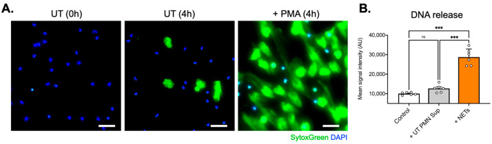
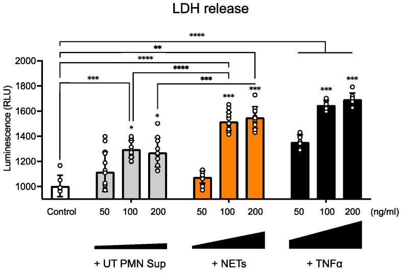
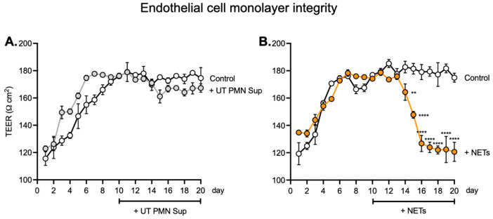
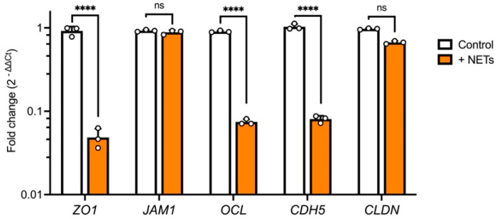
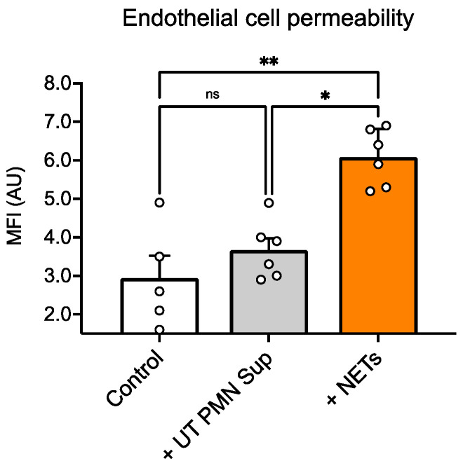
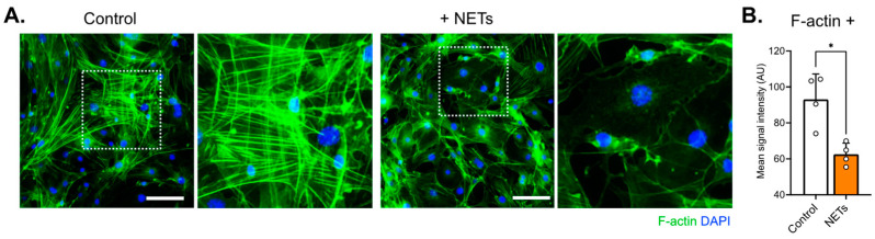
The integrity of the blood-labyrinth barrier (BLB) is essential for inner ear homeostasis, regulating the ionic composition of endolymph and perilymph and preventing harmful substance entry. Endothelial hyperpermeability, central in inflammatory and immune responses, is managed through complex intercellular communication and molecular signaling pathways. Recent studies link BLB permeability dysregulation to auditory pathologies like acoustic trauma, autoimmune inner ear diseases, and presbycusis. Polymorphonuclear granulocytes (PMNs), or neutrophils, significantly modulate vascular permeability, impacting endothelial barrier properties. Neutrophil extracellular traps (NETs) are involved in diseases with autoimmune and autoinflammatory bases. The present study evaluated the impact of NETs on a BLB cellular model using a Transwell® setup. Our findings revealed a concentration-dependent impact of NETs on human inner ear-derived endothelial cells. In particular, endothelial permeability markers increased, as indicated by reduced transepithelial electrical resistance, enhanced dextran permeability, and downregulated junctional gene expression (ZO1, OCL, and CDH5). Changes in cytoskeletal architecture were also observed. These preliminary results pave the way for further research into the potential involvement of NETs in BLB impairment and implications for auditory disorders.

Keywords: Meniere’s disease; TEER; blood–labyrinth barrier; hearing loss; tissue model.


Conflict of interest statement

The authors declare no conflicts of interest.

Figures

Figure 1
Figure 1
NET generation from healthy donors’ PMNs. (A) SytoxGreen and DAPI fluorescence captions depicting extracellular DNA and cell nuclei, respectively. (B) Spectro-photometrical determination of DNA quantities (NETs) in cell supernatants. Magnification: 10×; scale bars: 100 μm; ns: not significant; ***: p < 0.001; data are presented as the mean ± SD.
Figure 2
Figure 2
NETs induce endothelial toxicity in a dose-dependent manner. An LDH test was performed after 96 h of exposure to 50 ng/mL, 100 ng/mL, and 200 ng/mL of untreated PMN supernatant (UT PMN Sup) and NETs, with TNFα as a positive control. All three treatments showed increased luminescence intensity, indicating increased cellular toxicity compared with the control, with TNFα causing the most significant impact and the UT PMN supernatant having the lowest impact. At 100 ng/mL, NETs showed a significant increase in luminescence compared with both the EC medium-only control and the 100 ng/mL UT PMN supernatant. With the 200 ng/mL treatment, the cytotoxic luminescence signal increased significantly for NETs and TNFα, whereas UT PMN supernatant treatment values remained similar to values obtained with a 100 ng/mL concentration. Asterisks above the 100 ng/mL and 200 ng/mL bars indicate comparisons to the respective 50 ng/mL treatments. *: p < 0.05, **: p < 0.01, ***: p < 0.001, ****: p < 0.0001. Total number of individual treatment experiments (n = 3); data are presented as the mean ± SD.
Figure 3
Figure 3
TEER values are altered in the endothelial monolayer upon NET exposure. (A) TEER values after seeding human ECs on a Transwell® insert with a polyester membrane. Treatment with 100 ng/mL non-PMA-treated PMN supernatant (UT PMN Sup) started on day 10 and continued over the next 10 days. TEER measurements were performed once per day, with the first measurement starting 24 h after seeding and labeled as day 1. TEER measurement was performed in three consecutive repetitions for each individual well. Control cells were treated with media only. No significant difference between the UT PMN supernatant and control conditions was observed during the treatment period. (B) TEER values after treatment with NETs (100 ng/mL) vs. control showed a significant drop, starting from day 14 and continuing until the end of the treatment period. **: p < 0.01; ****: p < 0.0001; number of individual experiments with all treatment conditions (n = 6). Data are presented as the mean ± SD.
Figure 4
Figure 4
Expression of junctional genes is altered under the influence of NETs. Treatment with NETs (100 ng/mL) for 96 h resulted in a significant decrease in ZO1, OCL, and CDH5 expression, whereas JAM1 and CLDN levels remained unchanged compared with the control. Data were obtained from six donor-derived cell cultures and performed in triplicate (technical); number of individual experiments (n = 3); ns: not significant; ****: p < 0.0001; data are presented as the mean ± SD.
Figure 5
Figure 5
Increased EC permeability in the presence of NETs. Incubation of the endothelial cells with NETs (100 ng/mL) for 96 h produced a significant increase in dextran influx compared with the untreated cells, whereas the non-PMA-treated PMN supernatant (UT PMN Sup) did not result in any significant changes between control and treated cells. MFI: mean fluorescence intensity; ns: not significant; *: p < 0.05, **: p < 0.01; number of individual experiments with all treatment conditions (n = 6). Data are presented as the mean ± SD.
Figure 6
Figure 6
Decreased actin signal and minor rearrangement were observed in EC samples exposed to NETs. (A) Cells were stained with phalloidin conjugated with Alexa-488 (green) and DAPI (blue). In the NET-treated cells (100 ng/mL; right), changes in F-actin arrangement and a decrease in phalloidin intensity compared with control (left) were observed. Scale bar: 100 µm. (B) The fluorescence signals were weaker in NET-treated cells, as confirmed by signal quantification. *: p < 0.05, number of individual experiments (n = 3). Data are presented as the mean ± SD.

References

    1. Kumar P., Shen Q., Pivetti C.D., Lee E.S., Wu M.H., Yuan S.Y. Molecular mechanisms of endothelial hyperpermeability: Implications in inflammation. Expert Rev. Mol. Med. 2009;11:e19. doi: 10.1017/S1462399409001112. - DOI - PMC - PubMed
    1. Juhn S.K., Rybak L.P., Prado S. Nature of blood-labyrinth barrier in experimental conditions. Pt 1Ann. Otol. Rhinol. Laryngol. 1981;90:135–141. doi: 10.1177/000348948109000208. - DOI - PubMed
    1. Suzuki M., Kaga K. Development of blood-labyrinth barrier in the semicircular canal ampulla of the rat. Hear. Res. 1999;129:27–34. doi: 10.1016/S0378-5955(98)00214-7. - DOI - PubMed
    1. Juhn S.K., Rybak L.P., Fowlks W.L. Transport characteristics of the blood--perilymph barrier. Am. J. Otolaryngol. 1982;3:392–396. doi: 10.1016/S0196-0709(82)80016-1. - DOI - PubMed
    1. Shi X. Physiopathology of the cochlear microcirculation. Hear. Res. 2011;282:10–24. doi: 10.1016/j.heares.2011.08.006. - DOI - PMC - PubMed

LinkOut - more resources