. 2024 Sep 12;25(18):9854.
doi: 10.3390/ijms25189854.

Cell-Type-Specific Expression of Leptin Receptors in the Mouse Forebrain

Affiliations

Cell-Type-Specific Expression of Leptin Receptors in the Mouse Forebrain

Cade R Canepa et al. Int J Mol Sci. .

Abstract

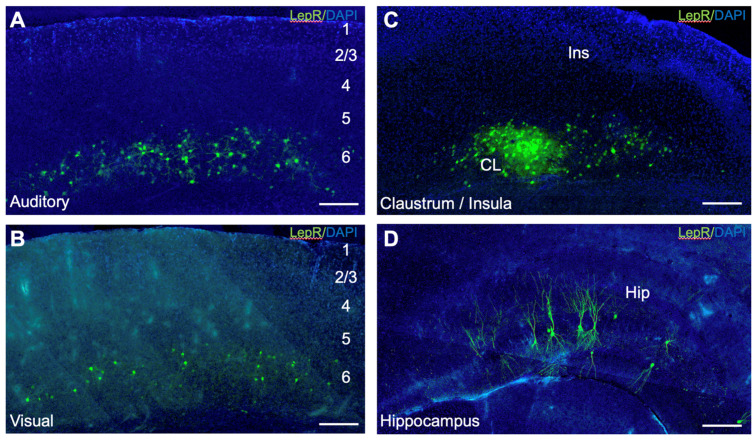
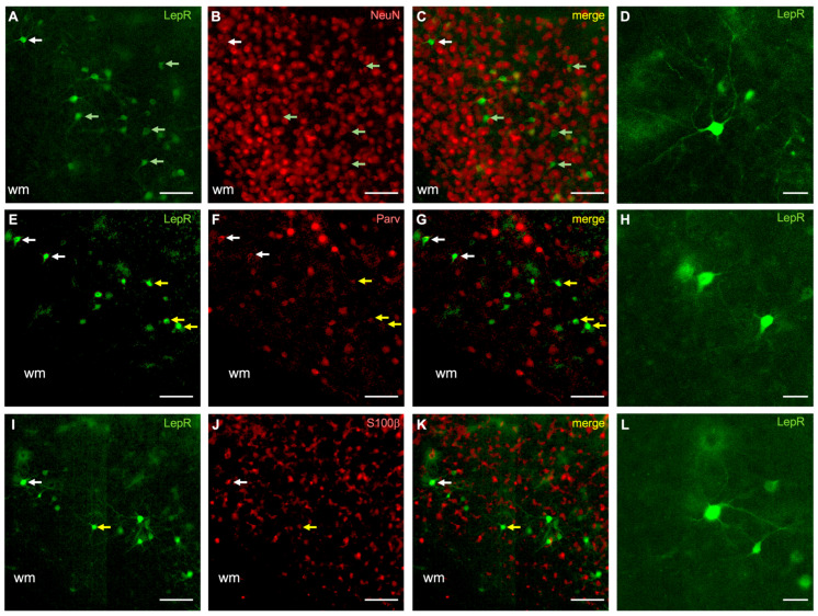
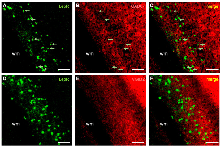
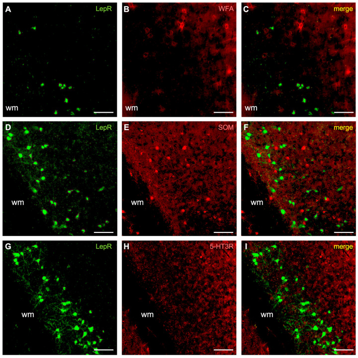
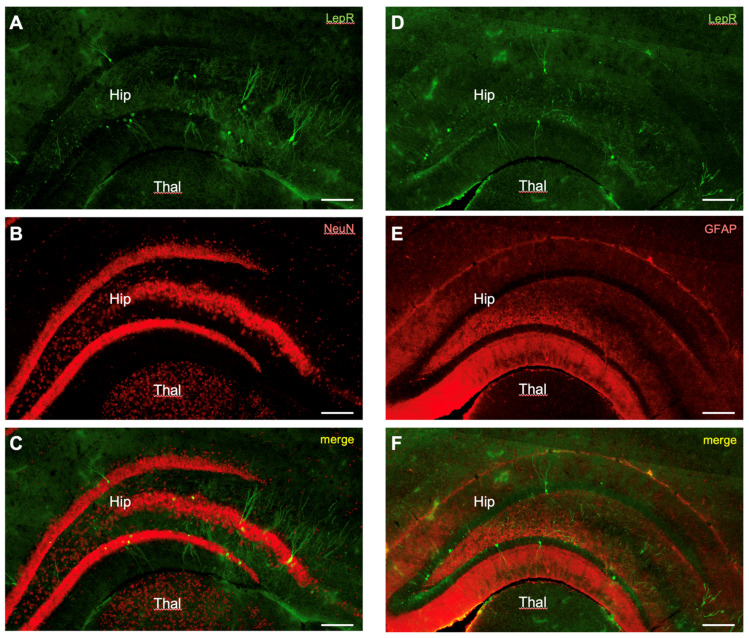
Leptin is a hormone produced by the small intestines and adipose tissue that promotes feelings of satiety. Leptin receptors (LepRs) are highly expressed in the hypothalamus, enabling central neural control of hunger. Interestingly, LepRs are also expressed in several other regions of the body and brain, notably in the cerebral cortex and hippocampus. These brain regions mediate higher-order sensory, motor, cognitive, and memory functions, which can be profoundly altered during periods of hunger and satiety. However, LepR expression in these regions has not been fully characterized on a cell-type-specific basis, which is necessary to begin assessing their potential functional impact. Consequently, we examined LepR expression on neurons and glia in the forebrain using a LepR-Cre transgenic mouse model. LepR-positive cells were identified using a 'floxed' viral cell-filling approach and co-labeling immunohistochemically for cell-type-specific markers, i.e., NeuN, VGlut2, GAD67, parvalbumin, somatostatin, 5-HT3R, WFA, S100β, and GFAP. In the cortex, LepR-positive cells were localized to lower layers (primarily layer 6) and exhibited non-pyramidal cellular morphologies. The majority of cortical LepR-positive cells were neurons, while the remainder were identified primarily as astrocytes or other glial cells. The majority of cortical LepR-positive neurons co-expressed parvalbumin, while none expressed somatostatin or 5-HT3R. In contrast, all hippocampal LepR-positive cells were neuronal, with none co-expressing GFAP. These data suggest that leptin can potentially influence neural processing in forebrain regions associated with sensation and limbic-related functions.

Keywords: auditory; claustrum; hippocampus; leptin; somatosensory; visual.


Conflict of interest statement

The authors declare no conflicts of interest.

Figures

Figure 1
Figure 1
Leptin receptor (LepR)-positive cells in the mouse forebrain. (A) Primary auditory cortex; (B) primary visual cortex; (C) insular cortex and claustrum; (D) hippocampus. Green labeling: LepR-positive cells. Blue labeling: DAPI counterstain. Scale bar: 200 μm.
Figure 2
Figure 2
LepR-positive cells in sensory cortices co-localize with NeuN, parvalbumin, and S100β. (AD) Neuronal marker (NeuN) co-staining; (EH) parvalbumin co-staining; (IL) S100β co-staining. In all panels, the green rendering indicates LepR-positive cells (A,E,I), the red rendering indicates neurochemical markers (B,F,J), and the yellow rendering illustrates double-labeling (C,G,K). The dendritic morphology of representative cells (D,H,L) is indicated by the white arrows in their respective rows. Light green arrows indicate non-double-labeled cells (AC). Yellow arrows indicate double-labeled cells (EG,IK). Scale bar in (G,H,L): 10 μm. Scale bar in other panels: 100 μm. wm: white matter.
Figure 3
Figure 3
Co-labeling of LepR-positive cells with GAD67 and VGlut2. (AC) Marker for GABAergic inhibitory neurons (GAD67). (DF) VGlut2 co-staining. The green rendering indicates LepR-positive cells (A,C,D,F), the red rendering indicates neurochemical markers (B,C,E,F), and the yellow rendering indicates double-labeling (C,F). Light green arrows indicate non-double-labeled cells (AC). Scale bar: 100 μm. wm: white matter.
Figure 4
Figure 4
LepR-positive cells co-stained for perineuronal nets (WFA), somatostatin (SOM) and serotonin receptor 3R (5-HT3R). (AC) Perineuronal net (WFA) co-staining. (DF) Somatostatin (SOM), calcium binding protein, co-staining. (GI) Co-staining for somatostatin (SOM) calcium binding protein. The green rendering depicts LepR-positive cells (A,D,G), the red rendering is for the neurochemical markers (B,E,H), and the yellow rendering illustrates double-labeling (C,F,I). Scale bar: 100 μm. wm: white matter.
Figure 5
Figure 5
LepR-positive cells in the hippocampus co-stained for NeuN and GFAP. (AC) Co-labeling of LepR-positive cells with NeuN. (DF) Co-labeling of LepR-positive cells for GFAP. The green rendering depicts LepR-positive cells (A,D), the red rendering is for the neurochemical marker (B,E), and the yellow rendering illustrates double-labeling (C,F). Scale bar: 100 μm. Hip: hippocampus. Thal: thalamus.

References

    1. Sørensen T.I., Echwald S., Holm J.C. Leptin in obesity. BMJ. 1996;313:953–954. doi: 10.1136/bmj.313.7063.953. - DOI - PMC - PubMed
    1. Schwartz M.W., Peskind E., Raskind M., Boyko E.J., Porte D. Cerebrospinal fluid leptin levels: Relationship to plasma levels and to adiposity in humans. Nat. Med. 1996;2:589–593. doi: 10.1038/nm0596-589. - DOI - PubMed
    1. Bacart J., Leloire A., Levoye A., Froguel P., Jockers R., Couturier C. Evidence for leptin receptor isoforms heteromerization at the cell surface. FEBS Lett. 2010;584:2213–2217. doi: 10.1016/j.febslet.2010.03.033. - DOI - PubMed
    1. Golden P.L., Maccagnan T.J., Pardridge W.M. Human blood-brain barrier leptin receptor. Binding and endocytosis in isolated human brain microvessels. J. Clin. Investig. 1997;99:14–18. doi: 10.1172/JCI119125. - DOI - PMC - PubMed
    1. Bjørbaek C., Elmquist J.K., Michl P., Ahima R.S., van Bueren A., McCall A.L., Flier J.S. Expression of leptin receptor isoforms in rat brain microvessels. Endocrinology. 1998;139:3485–3491. doi: 10.1210/endo.139.8.6154. - DOI - PubMed

LinkOut - more resources