. 2024 Sep 12;25(18):9863.
doi: 10.3390/ijms25189863.

Identification of a Novel Subset of Human Airway Epithelial Basal Stem Cells

Affiliations

Identification of a Novel Subset of Human Airway Epithelial Basal Stem Cells

Christopher Cheng et al. Int J Mol Sci. .

Abstract

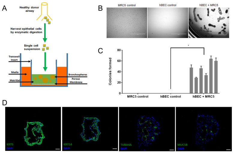
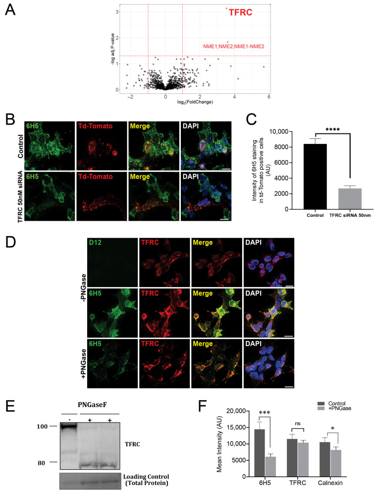
The basal cell maintains the airway's respiratory epithelium as the putative resident stem cell. Basal cells are known to self-renew and differentiate into airway ciliated and secretory cells. However, it is not clear if every basal cell functions as a stem cell. To address functional heterogeneity amongst the basal cell population, we developed a novel monoclonal antibody, HLO1-6H5, that identifies a subset of KRT5+ (cytokeratin 5) basal cells. We used HLO1-6H5 and other known basal cell-reactive reagents to isolate viable airway subsets from primary human airway epithelium by Fluorescence Activated Cell Sorting. Isolated primary cell subsets were assessed for the stem cell capabilities of self-renewal and differentiation in the bronchosphere assay, which revealed that bipotent stem cells were, at minimum 3-fold enriched in the HLO1-6H5+ cell subset. Crosslinking-mass spectrometry identified the HLO1-6H5 target as a glycosylated TFRC/CD71 (transferrin receptor) proteoform. The HLO1-6H5 antibody provides a valuable new tool for identifying and isolating a subset of primary human airway basal cells that are substantially enriched for bipotent stem/progenitor cells and reveals TFRC as a defining surface marker for this novel cell subset.

Keywords: CD71; aminooxy-sulfhydryl-biotin crosslinking; basal cells; bronchospheres; cell heterogeneity; fluorescence activated cell sorting; glycosylated transferrin receptor; monoclonal antibody; respiratory epithelium; stem cells.


Conflict of interest statement

The authors declare no conflicts of interest.

Figures

Figure 1
Figure 1
HLO1-6H5 identifies a subset of human basal cells. Representative immunohistochemical co-staining of human proximal airway with the HLO1-6H5 monoclonal antibody (mAb), in combination with canonical markers, that identify specific airway epithelial cell types such as (A) basal cells (KRT5, cytokeratin 5) and mitotically active basal cells (KRT14, cytokeratin 14). Green arrows indicate basal cells that are KRT5+/HLO1-6H5−. Red arrows indicate HLO1-6H5+ cells along the basal laminae that are KRT14-. HLO1-6H5 were also co-stained with markers for (B) luminal columnar cells (KRT8, cytokeratin 8), such as ciliated cells (TUBA4A, acetylated-alpha tubulin) and secretory cells (Muc5B, mucin 5B). (n = three donor samples) Scale bar: 20 µm. (C) Day 28 bronchospheres derived from human bronchial epithelial cells (hBECs) are also stained for basal cells (KRT5) and HLO1-6H5+ basal cells. Scale bar: 200 µm. (D) Representative flow cytometry plots of HLO1-6H5 co-expression with KRT5 or KRT14 in uncultured primary hBECs. (n = three donor samples).
Figure 2
Figure 2
Primary hBECs require MRC5 stromal cells to form bronchospheres composed of differentiated airway epithelial cell lineages. (A) Primary hBECs are harvested from proximal airway and seeded as single cells into MatriGel® matrix that is layered into a Transwell® insert to generate an air–liquid interface culture condition. (B) Bronchospheres grown for 14 days from unsorted hBECs co-cultured with MRC5 human fetal lung fibroblasts. Scale bars, 2000 µm. (C) Bar graph of number of bronchospheres formed by MRC5 fibroblasts, hBECs, and hBECs co-cultured with MRC5 fibroblasts across six donors, three technical replicates per donor. Data presented as mean (±S.D.). Paired t-test performed, * p < 0.05. (D) Bronchospheres grown for 42 days from unsorted hBECs form lumens in the interior of the spheroid and contain basal cells that are KRT5+, mitotically active basal cells (KRT14+), ciliated cells (TUBA4A+), and secretory goblet cells (MUC5B+). Scale bar: 50 µm.
Figure 3
Figure 3
Fluorescence activated cell sorting (FACS) -isolated HLO1-6H5+ basal cells are functionally enriched for a bipotent stem/progenitor cell population. (A) Representative data from FACS-isolation of HLO1-6H5 subsets for assessment of clonogenicity and potency in the bronchosphere assay (n = 5 donor samples). (B) After 14 days of culture, colonies were counted to determine which HLO1-6H5 subset was enriched for clonogenic cells. Scale bar: 2000 µm. (C) Scatter plot shows the number of colonies formed as a percentage relative to the number of input cells (colony-forming efficiency). Paired t-test performed, * p < 0.05. Data presented as mean (±S.D.). (D) Bronchospheres derived from HLO1-6H5+ basal cells were sectioned and analyzed by immunocytochemistry to determine if the cellular composition of the colonies included basal cells (KRT5), mitotically active basal cells (KRT14), ciliated cells (TUBA4A), and/or secretory cells (MUC5B). Scale bar: 50 µm.
Figure 4
Figure 4
HLO1-6H5+ basal cells are a subset of ITGA6+ (integrin alpha 6) airway epithelial cells and are distinct from NGFR+ (nerve growth factor receptor) basal cells in uncultured primary hBECs. (A) Representative flow cytometry plots of HLO1-6H5 combined with ITGA6 to FACS-isolate cell subsets for assessment of clonogenicity in the bronchosphere assay. (n = three donor samples) (B) At Day 14, colonies were imaged and counted to determine which sorted subsets were enriched for clonogenic cells. (C) Graphs of colony-forming efficiency of each HLO1-6H5/ITGA6 cell subset across three donors, three technical replicates per donor. (D) Representative flow cytometry plots of HLO1-6H5 combined with NGFR to FACS-isolate cell subsets for assessment of clonogenicity in the bronchosphere assay. (n = three donor samples). (E) At Day 14, colonies were imaged and counted to determine which sorted subsets were enriched for clonogenic cells. (F) Graphs of colony-forming efficiency of each HLO1-6H5/NGFR cell subset across three donors, three technical replicates per donor. Paired t-test was performed for each donor sample, * p < 0.05. n.s. = not statistically significant. Data presented as mean (±S.D.). Scale bar: 2000 µm.
Figure 5
Figure 5
The HLO1-6H5 antibody recognizes a glycosylated transferrin receptor (TFRC) protein. (A) Volcano plot comparing the difference in enrichment between the HLO1-6H5 mAb and isotype control in the ASB experiment. Proteins significantly enriched by the HLO1-6H5 mAb over the control (adjusted p-value < 0.05, log2 fold-change > 3.5) are highlighted in red. Number of independent replicates = 3. (B) Immunocytochemistry with HLO1-6H5 mAb (green) of HLO1-6H5+ HEK293 cells treated with control non-targeting siRNA, TFRC siRNA, and pcNDA3.1-myr-tdTomato to show HLO1-6H5 staining after TFRC knockdown (bottom panel) versus control (top panel). Note, cells positive for tdTomato indicate cells that receive siRNA as well. Scale bar = 20 μm. (C) Quantitation of intensity of HLO1-6H5 staining in cells positive for td-Tomato in both control and TFRC knocked down cells using multiple unpaired t-test where p < 0.0001 ****. (Number of independent replicates = 4). (D) Immunocytochemistry of HLO1-6H5+ HEK293 cells with HLO1-6H5 mAb (green), an isotype control (green), and commercially available anti-TFRC (red) to show HLO1-6H5 staining intensity and colocalization with TFRC in the absence (top two rows) and presence (bottom row) of PNGase F treatment. Scale bar = 20 µm. (E) Western blot analysis of PNGase-treated cell lysates to visualize shifts in TFRC band compared to untreated control. (F) Quantification of immunostaining intensities for HLO1-6H5, TFRC, and Calnexin (+ve control for PNGase F) in untreated and PNGase F treated cells using multiple unpaired t-test where p < 0.001 *** for HLO1-6H5 and p < 0.05 * for the positive control. Number of independent replicates = 2.

References

    1. Garcıá S.R., Deprez M., Lebrigand K., Cavard A., Paquet A., Arguel M.J., Magnone V., Truchi M., Caballero I., Leroy S., et al. Novel Dynamics of Human Mucociliary Differentiation Revealed by Single-Cell RNA Sequencing of Nasal Epithelial Cultures. Development. 2019;146:dev177428. doi: 10.1242/dev.177428. - DOI - PMC - PubMed
    1. Gomi K., Arbelaez V., Crystal R.G., Walters M.S. Activation of NOTCH1 or NOTCH3 Signaling Skews Human Airway Basal Cell Differentiation toward a Secretory Pathway. PLoS ONE. 2015;10:e0116507. doi: 10.1371/journal.pone.0116507. - DOI - PMC - PubMed
    1. Rock J.R., Rock J.R., Onaitis M.W., Onaitis M.W., Rawlins E.L., Rawlins E.L., Lu Y., Lu Y., Clark C.P., Clark C.P., et al. Basal Cells as Stem Cells of the Mouse Trachea and Human Airway Epithelium. Proc. Natl. Acad. Sci. USA. 2009;106:12771–12775. doi: 10.1073/pnas.0906850106. - DOI - PMC - PubMed
    1. Tadokoro T., Wang Y., Barak L.S., Bai Y., Randell S.H., Hogan B.L.M. IL-6/STAT3 Promotes Regeneration of Airway Ciliated Cells from Basal Stem Cells. Proc. Natl. Acad. Sci. USA. 2014;111:E3641–E3649. doi: 10.1073/pnas.1409781111. - DOI - PMC - PubMed
    1. Rock J.R., Gao X., Xue Y., Randell S.H., Kong Y.Y., Hogan B.L.M. Notch-Dependent Differentiation of Adult Airway Basal Stem Cells. Cell Stem Cell. 2011;8:639–648. doi: 10.1016/j.stem.2011.04.003. - DOI - PMC - PubMed