

Wall Shear Stress (WSS) Analysis in Atherosclerosis in Partial Ligated Apolipoprotein E Knockout Mouse Model through Computational Fluid Dynamics (CFD)
- PMID: 39337364
- PMCID: PMC11432177
- DOI: 10.3390/ijms25189877
Wall Shear Stress (WSS) Analysis in Atherosclerosis in Partial Ligated Apolipoprotein E Knockout Mouse Model through Computational Fluid Dynamics (CFD)
Abstract
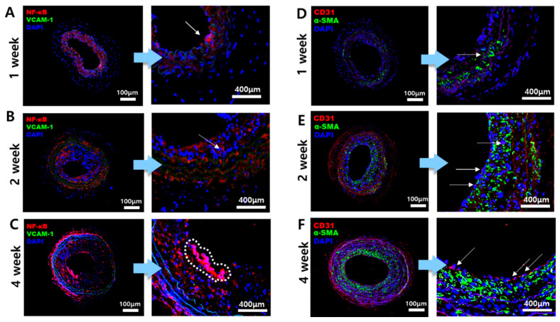
Atherosclerosis involves an inflammatory response due to plaque formation within the arteries, which can lead to ischemic stroke and heart disease. It is one of the leading causes of death worldwide, with various contributing factors such as hyperlipidemia, hypertension, obesity, diabetes, and smoking. Wall shear stress (WSS) is also known as a contributing factor of the formation of atherosclerotic plaques. Since the causes of atherosclerosis cannot be attributed to a single factor, clearly understanding the mechanisms and causes of its occurrence is crucial for preventing the disease and developing effective treatment strategies. To better understand atherosclerosis and define the correlation between various contributing factors, computational fluid dynamics (CFD) analysis is primarily used. CFD simulates WSS, the frictional force caused by blood flow on the vessel wall with various hemodynamic changes. Using apolipoprotein E knockout (ApoE-KO) mice subjected to partial ligation and a high-fat diet at 1-week, 2-week, and 4-week intervals as an atherosclerosis model, CFD analysis was conducted along with the reconstruction of carotid artery blood flow via magnetic resonance imaging (MRI) and compared to the inflammatory factors and pathological staining. In this experiment, a comparative analysis of the effects of high WSS and low WSS was conducted by comparing the standard deviation of time-averaged wall shear stress (TAWSS) at each point within the vessel wall. As a novel approach, the standard deviation of TAWSS within the vessel was analyzed with the staining results and pathological features. Since the onset of atherosclerosis cannot be explained by a single factor, the aim was to find the correlation between the thickness of atherosclerotic plaques and inflammatory factors through standard deviation analysis. As a result, the gap between low WSS and high WSS widened as the interval between weeks in the atherosclerosis mouse model increased. This finding not only linked the occurrence of atherosclerosis to WSS differences but also provided a connection to the causes of vulnerable plaques.
Keywords: ApoE-KO mice; atherosclerosis; computational fluid dynamics (CFD); plaque formation; standard deviation; wall shear stress (WSS).
Conflict of interest statement
The authors declare no conflicts of interest.
Figures


References
-
- Naghavi M., Wang H., Lozano R., Davis A., Liang X., Zhou M., Vollset S.E., Abbasoglu Ozgoren A., Abdalla S., Abd-Allah F., et al. Global, regional, and national age-sex specific all-cause and cause-specific mortality for 240 causes of death, 1990–2013: A systematic analysis for the Global Burden of Disease Study 2013. Lancet. 2015;385:117–171. doi: 10.1016/S0140-6736(14)61682-2. - DOI - PMC - PubMed
MeSH terms
Substances
LinkOut - more resources
Full Text Sources
Medical
Research Materials
Miscellaneous
