. 2024 Sep 13;25(18):9913.
doi: 10.3390/ijms25189913.

Quintessential Synergy: Concurrent Transient Administration of Integrated Stress Response Inhibitors and BACE1 and/or BACE2 Activators as the Optimal Therapeutic Strategy for Alzheimer's Disease

Affiliations

Quintessential Synergy: Concurrent Transient Administration of Integrated Stress Response Inhibitors and BACE1 and/or BACE2 Activators as the Optimal Therapeutic Strategy for Alzheimer's Disease

Vladimir Volloch et al. Int J Mol Sci. .

Abstract

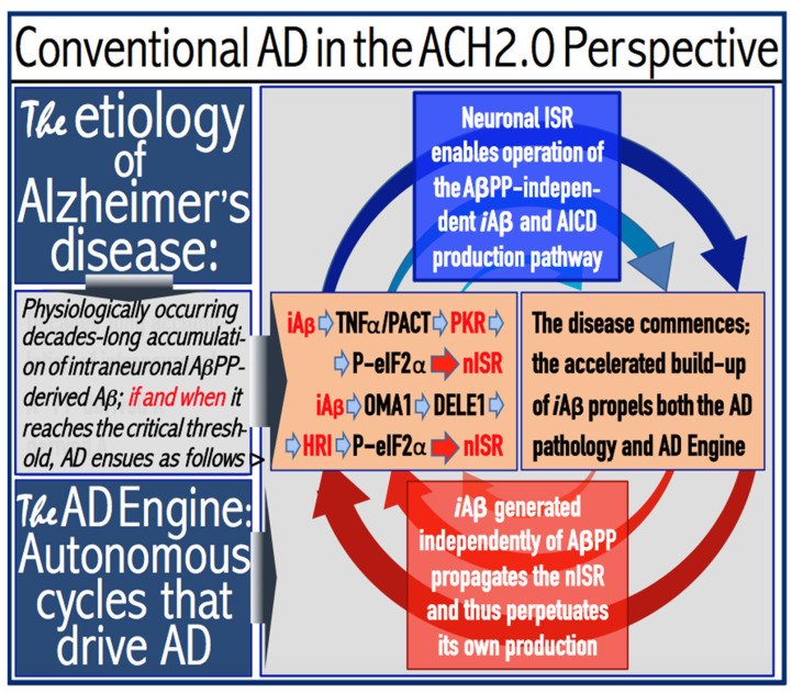
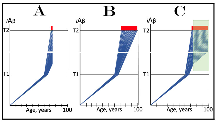
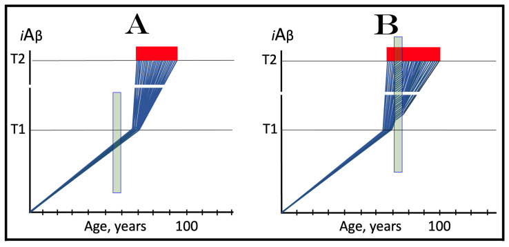
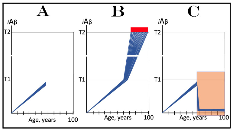
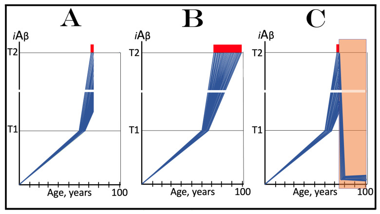
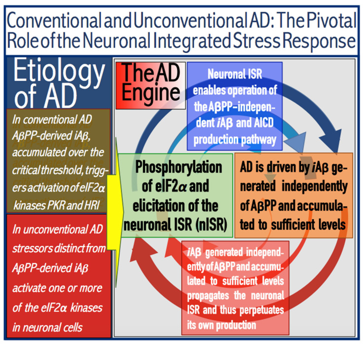
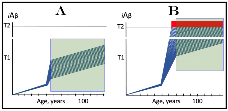
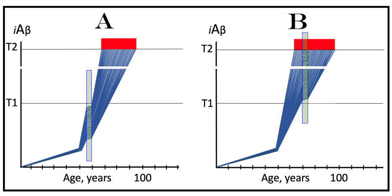
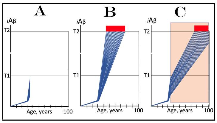
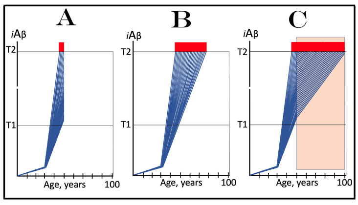
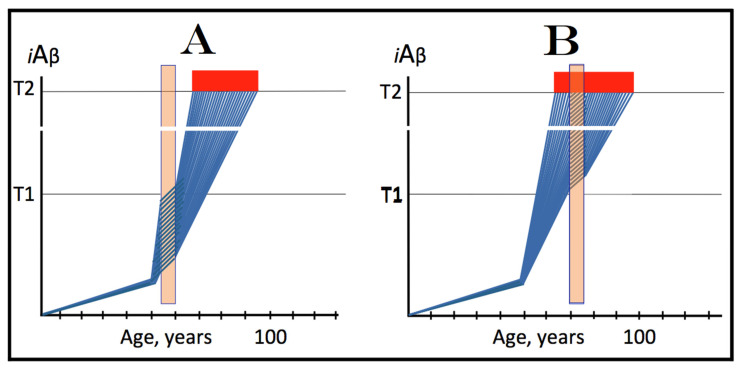
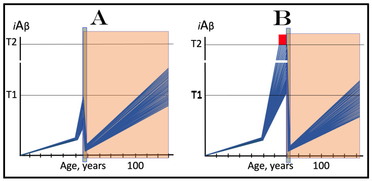
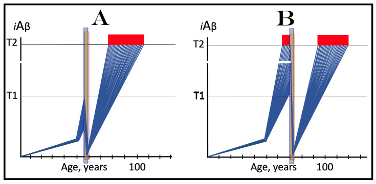
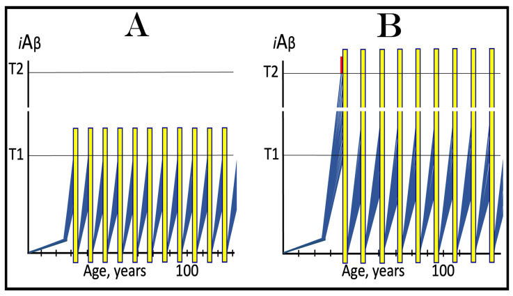
The present study analyzes two potential therapeutic approaches for Alzheimer's disease (AD). One is the suppression of the neuronal integrated stress response (ISR). Another is the targeted degradation of intraneuronal amyloid-beta (iAβ) via the activation of BACE1 (Beta-site Aβ-protein-precursor Cleaving Enzyme) and/or BACE2. Both approaches are rational. Both are promising. Both have substantial intrinsic limitations. However, when combined in a carefully orchestrated manner into a composite therapy they display a prototypical synergy and constitute the apparently optimal, potentially most effective therapeutic strategy for AD.

Keywords: AβPP-independent generation of iAβ; RNA-dependent asymmetric amplification of human AβPP mRNA; amyloid cascade hypothesis 2.0 (ACH2.0); conventional and unconventional Alzheimer’s disease; depletion of iAβ via the activation of BACE1 and/or BACE2; initiation of translation from the AUG codon encoding Met671 of the intact or 5′-truncated human AβPP mRNA; intraneuronal Aβ (iAβ); neuronal integrated stress response (ISR); suppression of the neuronal ISR; therapeutic strategies for conventional and unconventional Alzheimer’s disease.


Conflict of interest statement

The authors declare no conflicts of interest.

Figures

Figure 1
Figure 1
Dynamics of AβPP-derived iAβ accumulation in health and AD: rate of accumulation as a decisive variable. iAβ: intraneuronal Aβ. T1 threshold: levels of iAβ that trigger the activation of the eIF2α kinases PKR and/or HRI, elicitation of the neuronal ISR, and initiation of the AβPP-independent production of iAβ. T2 threshold: levels of iAβ, produced mainly in the AβPP-independent pathway, which trigger neuronal death. Blue lines: levels of iAβ in individual neurons of a person. Red Boxes: apoptotic zone; the fraction of neurons that have committed apoptosis (or necroptosis) or are dead. The rate of accumulation of AβPP-derived iAβ is the only variable in this figure; all other parameters are presumed constant. The occurrence of AD depends on a sufficient rate of accumulation of AβPP-derived iAβ and its timing is inversely proportional to the latter. Panel (A): the rate of accumulation of AβPP-derived iAβ is relatively high; the T1 threshold is crossed and, consequently, AD commences at about 65 years of age. Panels (B,C): the rate of accumulation of AβPP-derived iAβ decreases and the timing of the T1 crossing (and of the commencement of AD) increases; in panel (B), the crossing occurs and the disease commences at about 80 and in panel (C) at about 90 years of age. Panel (D): the rate of accumulation of AβPP-derived iAβ is such that neither is the T1 threshold crossed nor does AD occur within the lifetime of the individual. Note that with a sufficiently extended lifespan, both the T1 crossing and the occurrence of conventional AD would be inevitable at any rate of the accumulation of AβPP-derived iAβ.
Figure 2
Figure 2
The timing of AD and occurrence of AACD are functions of the extent of the T1 threshold. iAβ: intraneuronal Aβ. T0 threshold: levels of AβPP-derived iAβ that cause neuronal damage manifesting as AACD. T1 threshold: levels of iAβ that trigger the activation of the eIF2α kinases PKR and/or HRI, elicitation of the neuronal ISR, and initiation of the AβPP-independent production of iAβ. T2 threshold: levels of iAβ, produced mainly in the AβPP-independent pathway, which trigger neuronal death. Blue lines: levels of iAβ in individual neurons of a person. Red Boxes: apoptotic zone; the fraction of neurons that have committed apoptosis (or necroptosis) or are dead. Pink Boxes: zone of the occurrence of AACD; the condition commences with the crossing of the T0 threshold (provided it is lower that the T1 threshold) and morphs into AD following the T1 crossing. In this figure, the extent of the T1 threshold is the only variable; all other parameters (including the extent of the T0 threshold) are presumed constant. The occurrence of AD is a function of the extent of the T1 threshold and its timing is directly proportional to the latter. Panel (A): The extent of the T1 threshold is relatively low and below that of the T0 threshold. AD commences at about 65 years of age and no AACD occurs. Panel (B): The extent of the T1 threshold increases and is above that of the T0. AACD commences with the T0 crossing and morphs into AD when the T1 threshold is crossed. Panel (C): The extent of the T1 threshold increases further. AD commences later in life and the duration of AACD increases. Panel (D): The extent of the T1 threshold is such that neither is it crossed nor does AD occur within the lifespan of the individual. AACD, on the other hand, commences with the crossing of the T0 threshold and persists through the remaining lifetime.
Figure 3
Figure 3
Conventional Alzheimer’s disease: the ACH2.0 perspective. iAβ: intraneuronal Aβ. eIF2α: eukaryotic translation initiation factor 2 alpha. PKR and HRI: eIF2α–specific kinases. TNFα: tumor necrosis factor alpha; presumably, it activates the PKR kinase. PACT: activator of PKR. OMA1: mitochondrial dysfunction-activated mitochondrial protease. DELE1: mitochondrial substrate of OMA1; its cleavage results in the activation of HRI. nISR: neuronal integrated stress response. It is caused by phosphorylation of eIF2α and manifests in radical transcriptional and translational reprogramming; it provides “missing” components of the AβPP-independent iAβ generation pathway, thus activating it. AICD: AβPP Intracellular domain; one of the products of the processing of C99. AβPP-derived iAβ accumulates physiologically in a decades-long process. Conventional AD occurs only if and when AβPP-derived iAβ crosses the critical ISR-eliciting threshold. At this point, PKR and/or HRI are activated, eIF2α is phosphorylated, and the ISR is elicited. Under the ISR conditions, the crucial components of the AβPP-independent iAβ generation pathway are produced and the pathway is initiated. The entire Aβ output of this pathway is retained intraneuronally as iAβ. It rapidly accumulates and reaches AD pathology-causing levels. It is, therefore, the driver of the disease. It also sustains the activity of PKR and HRI, maintains eIF2α in a phosphorylated state, propagates the ISR conditions, and thus perpetuates its own production in the AβPP-independent pathway, thus propelling the progression of AD. The repeated cycles of these activities, symbolized in the figure by the arched red and blue arrows, comprise the engine that drives AD, the “AD Engine”. Note that the increase in iAβ produced independently of AβPP is accompanied by a proportional increase in levels of AICD. The AβPP intracellular domain was shown to be competent of interfering with AD-associated processes but its potential contribution to the disease remains to be determined [141,142,143,144,145,146,147,148,149,150,151,152,153,154,155,156].
Figure 4
Figure 4
Effect of suppression of the neuronal ISR in the prevention of conventional AD. iAβ: intraneuronal Aβ. T1 threshold: levels of iAβ that trigger the activation of the eIF2α kinases PKR and/or HRI, elicitation of the neuronal ISR, and initiation of the AβPP-independent production of iAβ. T2 threshold: levels of iAβ, produced mainly in the AβPP-independent pathway, which trigger neuronal death. Blue lines: levels of iAβ in individual neurons of a person. Red Box: apoptotic zone; the fraction of neurons that have committed apoptosis (or necroptosis) or are dead. Green Box: the duration of the presence of an inhibitor of the IRS. Panel (A): The initial state of the levels of AβPP-derived iAβ in individual neurons of a healthy person. They are all sub-T1. Panel (B): Evolution of the initial state in the absence of the drug. AβPP-derived iAβ reaches and crosses the T1 threshold in all affected neurons. The neuronal ISR is elicited and the AβPP-independent iAβ generation pathway activated. Its iAβ product rapidly accumulates, reaches AD pathology-causing levels, and eventually crosses the T2 threshold. As more and more neurons commit apoptosis, the disease enters its end stage. Panel (C): Evolution of the initial state in the presence of an ISR-inhibiting drug. The effect manifests only following the T1 crossing. The elicitation of the ISR is suppressed. The AβPP-independent iAβ generation pathway remains inoperative. The accumulation of AβPP-derived iAβ continues at the pre-T1 crossing rate. It does not reach AD pathology-causing levels and AD symptoms do not occur for the duration of the treatment.
Figure 5
Figure 5
Effect of inhibition of the neuronal ISR in the treatment of conventional AD. iAβ: intraneuronal Aβ. T1 threshold: levels of iAβ that trigger the activation of the eIF2α kinases PKR and/or HRI, elicitation of the neuronal ISR, and initiation of the AβPP-independent production of iAβ. T2 threshold: levels of iAβ, produced mainly in the AβPP-independent pathway, which trigger neuronal death. Blue lines: levels of iAβ in individual neurons. Red Box: apoptotic zone; the fraction of neurons that have committed apoptosis (or necroptosis) or are dead. Green Box: the duration of the presence of an inhibitor of the IRS. Panel (A): The initial state of the levels of AβPP-derived iAβ in individual neurons of a healthy person. In all affected neurons, iAβ has crossed the T1 threshold, the integrated stress response has been elicited, and the AβPP-independent iAβ production pathway has been activated. In a fraction of the neurons, iAβ has crossed the T2 threshold and AD symptoms have manifested. Panel (B): Evolution of the initial state in the absence of the drug. iAβ, produced mainly independently of AβPP, continues its accumulation unimpeded. Eventually, it crosses the T2 threshold in a sufficient fraction of the neurons and the disease enters its end stage. Panel (C): Evolution of the initial state in the presence of an ISR-inhibiting drug. With the suppression of the integrated stress response, the supply of components essential for the operation of the AβPP-independent iAβ production pathway ceases and the pathway is disabled. At this point, the neurons that already crossed the T2 threshold are unredeemable, and in a large fraction of the remaining neurons iAβ has reached AD pathology-causing levels. Moreover, the accumulation of AβPP-derived iAβ in the viable neurons continues but at the slow, pre-T1 crossing rate. The progression of the disease would not be stopped but rather would continue at a reduced rate for the duration of the treatment.
Figure 6
Figure 6
Effect of transient inhibition of the neuronal ISR in the prevention and treatment of conventional AD. iAβ: intraneuronal Aβ. T1 threshold: levels of iAβ that trigger the activation of the eIF2α kinases PKR and/or HRI, elicitation of the neuronal ISR, and initiation of the AβPP-independent production of iAβ. T2 threshold: levels of iAβ, produced mainly in the AβPP-independent pathway, which trigger neuronal death. Blue lines: levels of iAβ in individual neurons of a person. Red Box: apoptotic zone; the fraction of neurons that have committed apoptosis (or necroptosis) or are dead. Green Boxes: the duration of the presence of an inhibitor of the IRS. Panel (A): The transient treatment with the ISR-inhibiting drug is administered prior to the T1 crossing by AβPP-derived iAβ. At this stage, the ISR has not yet been elicited and the administration of its inhibitors serves no purpose and does not affect the continuous accumulation of AβPP-derived iAβ. When the T1 threshold is crossed, the ISR is elicited, the AβPP-independent iAβ production pathway is activated, and AD progresses unimpeded. Panel (B): The transient ISR suppression treatment is administered when the AβPP-independent iAβ generation pathway is operational and depends on the occurrence of the ISR. Inhibition of the latter disables the former. iAβ continues to accumulate for the duration of the treatment due to the influx of the AβPP-derived iAβ. This accumulation occurs at the slow pre-T1 crossing rate and so does the progression of the disease. When the drug is withdrawn, the ISR is re-elicited (due to over-T1 levels of iAβ) and the AβPP-independent iAβ production pathway re-activated. Both the iAβ accumulation and the progression of AD resume at the fast pre-treatment rate. The transient ISR suppression provides only a short reprieve of slow progression of AD lasting no more than the duration of the treatment.
Figure 7
Figure 7
Effect of long-term iAβ degradation therapy in the prevention of conventional AD. iAβ: intraneuronal Aβ. T1 threshold: levels of iAβ that trigger the activation of the eIF2α kinases PKR and/or HRI, elicitation of the neuronal ISR, and initiation of the AβPP-independent production of iAβ. T2 threshold: levels of iAβ, produced mainly in the AβPP-independent pathway, which trigger neuronal death. Blue lines: levels of iAβ in individual neurons of a person. Red Box: apoptotic zone; the fraction of neurons that have committed apoptosis or are dead. Orange Box: the duration of the presence of activators of BACE1 and/or BACE2. Panel (A): The initial state of the levels of AβPP-derived iAβ in individual neurons of a healthy individual. In all neurons, these levels are below the T1 threshold. Panel (B): Evolution of the initial state in the absence of the treatment. AβPP-derived iAβ reaches and crosses the T1 threshold in all affected neurons. The PKR and/or HRI kinases are activated, eIF2α phosphorylated, the neuronal ISR elicited, and the AβPP-independent iAβ generation pathway initiated. Its iAβ product rapidly accumulates, reaches AD pathology-causing levels, and eventually crosses the T2 threshold. As more and more neurons commit apoptosis, the disease enters its end stage. Panel (C): Evolution of the initial state in the presence of a drug that activates BACE1 and/or BACE2. iAβ is rapidly depleted. Its levels represent equilibrium between its degradation and the continuous influx of AβPP-derived iAβ and are maintained at a low level for the duration of the treatment. The T1 threshold will not be reached, and conventional AD will not occur in the treated individual.
Figure 8
Figure 8
Effect of long-term iAβ degradation therapy in the treatment of conventional AD. iAβ: intraneuronal Aβ. T1 threshold: levels of iAβ that trigger the activation of the eIF2α kinases PKR and/or HRI, elicitation of the neuronal ISR, and initiation of the AβPP-independent production of iAβ. T2 threshold: levels of iAβ, produced mainly in the AβPP-independent pathway, which trigger neuronal death. Blue lines: levels of iAβ in individual neurons of a person. Red Box: apoptotic zone; the fraction of neurons that have committed apoptosis (or necroptosis) or are dead. Orange Box: the duration of the presence of activators of BACE1 and/or BACE2. Panel (A): The initial state of the levels of AβPP-derived iAβ in individual neurons of a healthy person. In all affected neurons, iAβ has crossed the T1 threshold, the integrated stress response has been elicited, and the AβPP-independent iAβ production pathway has been activated. In a fraction of the neurons, iAβ has crossed the T2 threshold and AD symptoms have manifested. Panel (B): Evolution of the initial state in the absence of the drug. The accumulation of iAβ, produced predominantly independently in the AβPP-independent pathway, continues unimpeded. Eventually, it crosses the T2 threshold in a sufficient fraction of the neurons and the disease enters its end stage. Panel (C): Evolution of the initial state in the presence of the BACE1- and/or BACE2-activating drug. iAβ is being degraded by the drug in the viable neurons and its levels rapidly decrease (provided that activated BACE1 and/or BACE2 are capable of such efficient iAβ degradation; see Section 23 below). When they are below the T1 threshold, the ISR conditions cease and the AβPP-independent iAβ generation pathway is rendered inoperative. The levels of iAβ decrease further and are maintained at a low equilibrium with the influx of AβPP-derived iAβ by the drug. Neither is the T1 threshold reached nor does AD resume for the duration of the treatment.
Figure 9
Figure 9
Effects of the transient activation of BACE1 and/or BACE2 in the prevention and treatment of conventional AD. iAβ: intraneuronal Aβ. T1 threshold: levels of iAβ that trigger the activation of the eIF2α kinases PKR and/or HRI, elicitation of the neuronal ISR and initiation of the AβPP-independent production of iAβ. T2 threshold: levels of iAβ, produced mainly in the AβPP-independent pathway, which trigger neuronal death. Blue lines: levels of iAβ in individual neurons. Red Box: apoptotic zone; the fraction of neurons that have committed apoptosis (or necroptosis) or are dead. Orange Box: the duration of the presence of activators of BACE1 and/or BACE2. Panel (A): The transient treatment with the BACE1- and/or BACE2-activating drug is administered prior to the T1 crossing by AβPP-derived iAβ. Following the activation of BACE1 and/or BACE2, iAβ is rapidly depleted. When the drug is withdrawn, the de novo accumulation of AβPP-derived iAβ resumes from a low baseline at the pre-treatment rate. Its levels would not reach the T1 threshold and conventional AD would not occur within the lifetime of the treated individual; Panel (B): The transient treatment with the BACE1- and/or BACE2-activating drug is administered to a symptomatic AD patient. At the time of the treatment, iAβ has crossed the T1 threshold in all affected neurons. The ISR has been elicited and the AβPP-independent iAβ generation pathway activated. Its iAβ product has rapidly accumulated, reached AD pathology-causing levels, crossed the T2 threshold in a fraction the neurons, and AD symptoms have manifested. Provided the activated BACE1 and/or BACE2 are sufficiently efficient in the degradation of iAβ (see Section 23 below), its levels are rapidly reduced. When the drug is withdrawn, the AβPP-independent iAβ production pathway is no longer operational. The de novo accumulation of AβPP-derived iAβ resumes from a low baseline at the pre T1-crossing rate. The T1 threshold would not be reached and AD would not recur within the lifetime of the treated patient.
Figure 10
Figure 10
Principal stages of mammalian RNA-dependent mRNA amplification. Boxed line: sense RNA. Single line: antisense RNA. “AUG”: translation-initiating codon. “TCE”: 3′-terminal complementary element of the antisense RNA; “ICE”: internal complementary element of the antisense RNA. Yellow circle: enzymatic complex comprised of helicase, nucleotide-modifying activity, and nucleotide-cleaving activity. Blue lines: RNA molecules following their separation by the helicase-containing complex. Red arrows: location of the cleavage of the chimeric RNA intermediate by the helicase complex. (Top panel): conventionally genome-transcribed mRNA serves as the “progenitor” in the mRNA amplification process. (Middle panel): Principal stages of the “chimeric” pathway of RNA-dependent amplification of mammalian mRNA. Stage 1: RdRp transcribes antisense complement from the progenitor mRNA template. Stage 2: Separation of sense and antisense RNA. The helicase complex binds the 3′-terminal poly(A) component of the double-stranded structure and moves along the sense strand separating it from antisense RNA and modifying about every fifth nucleotide. Stage 3: separated antisense RNA folds into a self-priming structure; TCE/ICE interaction is essential in this process. Stage 4: self-primed antisense RNA is extended by RdRp; the resulting product forms a hairpin-like structure. Stage 5: Complementary sense and antisense components of the hairpin-like structure are separated by the helicase complex. The helicase complex commences at the 3′-terminal poly(A) of the sense RNA; it introduces nucleotide modifications that presumably prevent the RNA strands from re-annealing. Stage 6: when the single-stranded portion of the hairpin-like structure is reached, the helicase complex cleaves the chimeric RNA intermediate. Stage 7: End products of the chimeric mRNA amplification pathway. Importantly, the ICE element is located within a segment of the antisense RNA corresponding to the 5′UTR of the mRNA progenitor; consequently, the chimeric RNA end product contains the intact coding region of the progenitor mRNA. (Bottom panel): The ICE is situated within a segment of the antisense RNA corresponding to the coding region of the mRNA progenitor. Accordingly, the chimeric mRNA end product contains only a 3′-portion of the coding region of the mRNA progenitor. In this scenario, the translational outcome is determined by the location of the first translation initiation codon. If it were within the remaining segment of the coding region and in-frame with the protein-coding nucleotide sequence, translation would yield the C-terminal fragment of the polypeptide encoded by the progenitor mRNA. Stages 3′7′ correspond to stages 37.
Figure 11
Figure 11
Human AβPP mRNA is a potential progenitor in the asymmetric RNA-dependent amplification process; the resulting chimeric mRNA end product encodes the C100 fragment of AβPP. Small letters: nucleotide sequences of the relevant segments of the antisense human AβPP RNA. Capital letters: nucleotide sequences of the sense human AβPP mRNA segments resulting from the extension of self-primed antisense RNA. Letters highlighted in yellow: the TCE (top) and the ICE (bottom) components of the human antisense AβPP RNA. “2011–2013”: nucleotide locations (counted from the 3′ end of the antisense AβPP RNA) of the “uac” (highlighted in blue), the complement of the “AUG” (highlighted in green) encoding Met671 in the human AβPP mRNA. Panels (ac) correspond to Stages 3′, 4′, and 6′ of Figure 10. Panel (a): human antisense AβPP RNA folds into self-priming structure; TCE/ICE interaction is essential in this process. Panel (b): self-primed human antisense AβPP RNA is extended into a segment (highlighted in gray) of the human sense AβPP mRNA. Red arrow: when the helicase complex reaches the single-stranded portion of the chimeric hairpin-like structure, the cleavage occurs either at the 3′ end of the loop (shown) or at a mismatch within the TCE/ICE segment. Panel (c): The chimeric mRNA end product of RNA-dependent amplification of human AβPP mRNA (highlighted in gray). It consists of the antisense segment covalently attached to severely 5′-truncated AβPP mRNA. It retains only the 3′ portion of the coding region of the progenitor mRNA. Its first functional translation initiation codon is the AUG encoding Met671. Its translation would result in the C100 fragment of AβPP generated independently of the latter.
Figure 12
Figure 12
Projected effect of targeted iAβ degradation via the activation of BACE1 and/or BACE2 in the treatment of conventional symptomatic AD. iAβ: intraneuronal Aβ. T1 threshold: levels of iAβ that trigger the activation of the eIF2α kinases PKR and/or HRI, elicitation of the neuronal ISR, and initiation of the AβPP-independent production of iAβ. T2 threshold: levels of iAβ, produced mainly in the AβPP-independent pathway, which trigger neuronal death. Blue lines: levels of iAβ in individual neurons of a person. Red Box: apoptotic zone; the fraction of neurons that have committed apoptosis (or necroptosis) or are dead. Orange Box: the duration of the presence of activators of BACE1 and/or BACE2. Panel (A): The initial state of the levels of AβPP-derived iAβ in individual neurons of a healthy person. In all affected neurons, iAβ has crossed the T1 threshold, the integrated stress response has been elicited, and the AβPP-independent iAβ production pathway has been activated. In a fraction of the neurons, iAβ has crossed the T2 threshold and AD symptoms have manifested. Panel (B): Evolution of the initial state in the absence of the drug. The accumulation of iAβ, produced mainly independently of AβPP continues unimpeded. Eventually it crosses the T2 threshold in a sufficient fraction of the neurons and the disease enters its end stage. Panel (C): Evolution of the initial state in the presence of a BACE1- and/or BACE2-activating drug. The rate of degradation of iAβ by activated BACE1 and/or BACE2 does not match that of the influx of iAβ produced in the AβPP-independent pathway. The accumulation of the latter continues although at a reduced rate. The progression of the disease is not stopped but is merely slowed down for the duration of the treatment.
Figure 13
Figure 13
Composite therapy for conventional symptomatic AD: transient, ISR inhibitor-mediated suppression of the AβPP-independent production of iAβ for the duration of its concurrent, BACE activator-mediated depletion below the T1 threshold. iAβ: intraneuronal Aβ. T1 threshold: levels of iAβ that trigger the activation of the eIF2α kinases PKR and/or HRI, elicitation of the neuronal ISR, and initiation of the AβPP-independent production of iAβ. T2 threshold: levels of iAβ, produced mainly in the AβPP-independent pathway, which trigger neuronal death. Blue lines: levels of iAβ in individual neurons of a person. Red Box: apoptotic zone; the fraction of neurons that have committed apoptosis (or necroptosis) or are dead. Orange Boxes: the duration of the presence of activators of BACE1 and/or BACE2. In both panels, by the time the composite therapy commences, AβPP-derived iAβ has crossed the T1 in all affected neurons of an AD patient, the ISR has been elicited, and the AβPP-independent iAβ production pathway has been rendered operational; iAβ has rapidly accumulated, its levels have crossed the T2 threshold in a fraction of the neurons, and AD symptoms have manifested. Panel (A): The transient administration of an ISR inhibitor overlaps concurrently with the long-term treatment with BACE activators. With the neuronal ISR suppressed, the AβPP-independent iAβ generation pathway is disabled and the influx of its iAβ product ceases. Activated BACE1 and/or BACE2 efficiently and rapidly deplete iAβ to below-T1 levels. At this point, the ISR inhibitor can be withdrawn and the AβPP-independent iAβ production pathway remains inoperative. The depletion of iAβ continues until its levels reach equilibrium with the influx of AβPP-derived iAβ. The levels of iAβ would not be restored to the T1 threshold, the ISR would not be re-elicited, the AβPP-independent iAβ production pathway would not be re-activated, and AD would not recur in the presence of BACE activators. Panel (B): Similar to panel A except for the duration of treatment with BACE activators. In this panel ISR inhibitors and BACE activators are administered concurrently and transiently. The ISR-inhibiting drug is withdrawn when iAβ is depleted to below-T1 levels, and BACE1- and/or BACE2-activating drugs are removed when iAβ is depleted to the desired low levels. Following the composite therapy, the accumulation of AβPP-derived iAβ resumes de novo, from a low baseline and at the slow pre-T1 crossing rate. It will not reach the T1 threshold and AD will not recur within the remaining lifetime of the treated patient.
Figure 14
Figure 14
Unconventional AD is caused by the neuronal ISR elicited by stressors distinct from AβPP-derived iAβ. intraneuronal Aβ. eIF2α: eukaryotic translation initiation factor 2 alpha. PKR and HRI: eIF2α–specific kinases. nISR: neuronal integrated stress response. It is caused by phosphorylation of eIF2α at its Ser51 and manifests in radical transcriptional and translational reprogramming; it provides “missing” components of the AβPP-independent iAβ generation pathway thus activating it. AICD: AβPP intracellular domain; one of the products of the gamma-cleavage. The AD Engine: escalating cycles of self-stimulated production of iAβ in the AβPP-independent pathway (symbolized by arched red and blue arrows). In conventional AD, the activation of the PKR and HRI kinases in neuronal cells is triggered by AβPP-derived iAβ accumulated over the T1 threshold “conventional stressor”). In unconventional AD, the activation of the eIF2α kinases in the neurons is accomplished by stressors distinct from AβPP-derived iAβ (“unconventional stressors”). Diverse pathways leading to conventional and unconventional forms of AD conflate at the stage of the phosphorylation of eIF2α. The ensuing elicitation of the neuronal ISR is the pivotal point and realizes the uniform function in both conventional and unconventional forms of AD: it supplies the essential components of the AβPP-independent iAβ generation pathway and thus enables its operation. iAβ produced in this pathway fulfills two functions. One, it drives AD pathology and, two, it sustains the activity of PKR and HRI, maintains eIF2α in the phosphorylated state, propagates the ISR conditions, and thus perpetuates its own production in the AβPP-independent pathway, thus propelling the progression of AD. Note that the activation of the AβPP-independent iAβ generation pathway equates with the commencement of AD in conventional but not unconventional cases of the disease. AD (both conventional and unconventional) commences when the AβPP-independent iAβ production pathway becomes self-sustainable, i.e., the levels of iAβ are above the T1 threshold. In conventional AD, this self-sustainability is attained from the instance of the activation of the pathway, which is triggered by the over-T1 levels of AβPP-derived iAβ. In unconventional AD, the AβPP-independent iAβ generation pathway is activated below the T1 threshold and the disease is triggered only when its iAβ product accumulates to sufficient (i.e., above T1) levels. Hence, “…and accumulated to sufficient levels…” stated in the figure in relation to iAβ generated independently of AβPP is relevant to the cases of unconventional AD only; in conventional AD, the AβPP-independent production of iAβ (and the disease) initiates when the levels of AβPP-derived iAβ are already sufficiently, over-T1, high (see Figure 3).
Figure 15
Figure 15
Dynamics of iAβ accumulation in unconventional AD: Effect of long-term unconventional activation of the AβPP-independent iAβ generation pathway. iAβ: intraneuronal Aβ. T1 threshold: levels of iAβ that trigger the activation of the eIF2α kinases PKR and/or HRI, elicitation of the neuronal ISR and initiation of the AβPP-independent production of iAβ. T2 threshold: levels of iAβ, produced mainly in the AβPP-independent pathway, which trigger neuronal death. Blue lines: levels of iAβ in individual neurons of a person. Red Box: apoptotic zone; the fraction of neurons that have committed apoptosis (or necroptosis) or are dead. Pink Box: duration of the occurrence of unconventional stressors capable of eliciting the neuronal ISR. Panel (A): Dynamics of accumulation of AβPP-derived iAβ in a healthy individual. iAβ would not reach the T1 threshold and AD would not occur unless the neuronal ISR is elicited by an unconventional stressor and, consequently, the AβPP-independent iAβ production pathway is activated unconventionally. Panel (B): An event occurs or a condition develops that cause the long-term occurrence of unconventional stressors. The neuronal ISR is elicited and the AβPP-independent iAβ generation pathway is activated unconventionally and is sustained by a life-long presence of unconventional stressors. iAβ produced in this pathway rapidly accumulates, crosses the T1 threshold, renders the pathway self-sustainable, and unconventional AD ensues.
Figure 16
Figure 16
Dynamics of iAβ accumulation in unconventional AD: effect of transient unconventional activation of the AβPP-independent iAβ generation pathway. iAβ: intraneuronal Aβ. T1 threshold: levels of iAβ that trigger the activation of the eIF2α kinases PKR and/or HRI, and elicitation of the neuronal ISR. T2 threshold: levels of iAβ which trigger neuronal death. Blue lines: levels of iAβ in individual neurons. Red Box: apoptotic zone; the fraction of neurons that have committed apoptosis or are dead. Pink Boxes: duration of the occurrence of unconventional stressors capable of eliciting the neuronal ISR and of unconventionally activating the AβPP-independent iAβ production pathway. Panel (A): The duration of the occurrence of unconventional stressors is insufficient to cause the T1 crossing within the lifetime of the individual. Unconventional stressors elicit the neuronal ISR, unconventionally activate the AβPP-independent iAβ generation pathway, and its iAβ product rapidly accumulates. When unconventional stressors are no longer present, operation of the AβPP-independent iAβ production pathway ceases. AβPP-derived iAβ resumes its accumulation at a pre-unconventional stressors occurrence rate. It does not reach the T1 threshold and no AD occurs within the lifetime of the individual. Panel (B): The duration of the occurrence of unconventional stressors is sufficient for iAβ produced in the unconventionally activated AβPP-independent iAβ generation pathway to cross the T1 threshold in all neurons. Following the T1 crossing, this pathway becomes self-sustainable. The withdrawal of unconventional stressors at this stage would affect neither the operation of the AβPP-independent iAβ production pathway nor the progression of AD. Panel (C): At the time of withdrawal of unconventional stressors, iAβ crossed the T1 in only a fraction of the neurons where the AβPP-independent iAβ production pathway remains operational and the AD pathology progresses unimpeded. In sub-T1 neurons, the AβPP-independent iAβ production pathway is rendered inoperative but the accumulation of AβPP-derived iAβ continues. When it crosses the T1 threshold, the AβPP-independent iAβ production pathway is activated and the progression of the AD pathology commences.
Figure 17
Figure 17
Transient unconventional activation of the AβPP-independent iAβ generation pathway may cause unconventional AD in a delayed manner. iAβ: intraneuronal Aβ. T1 threshold: levels of iAβ that trigger the activation of the eIF2α kinases PKR and/or HRI, elicitation of the neuronal ISR, and initiation of the AβPP-independent production of iAβ. T2 threshold: levels of iAβ, produced mainly in the AβPP-independent pathway, which trigger neuronal death. Blue lines: levels of iAβ in individual neurons of a person. Red Box: apoptotic zone; the fraction of neurons that have committed apoptosis (or necroptosis) or are dead. Pink Boxes: duration of the occurrence of unconventional stressors capable of eliciting the neuronal ISR and, consequently, of unconventionally activating the AβPP-independent iAβ production pathway. Panel (A): The delayed effect of a single transient unconventional activation of the AβPP-independent iAβ production pathway. The transient occurrence of unconventional stressors elicits the neuronal ISR and unconventionally activates the AβPP-independent iAβ generation pathway. Its operation ceases when stressors are no longer present; its transiently produced iAβ product has accumulated to a substantial extent but its levels are still below the T1 threshold. The accumulation of AβPP-derived iAβ resumes at a slow, pre-unconventional stressors occurrence rate but from a significantly elevated baseline. Consequently, the T1 crossing occurs much sooner than it would in the absence of the unconventional activity of the AβPP-independent iAβ generation pathway. At this point, the pathway is re-activated and sustained conventionally by over-T1 levels of iAβ, and AD ensues. Panel (B): The delayed effect of the recurrent rounds of the unconventional transient activity of the AβPP-independent iAβ generation pathway. The transient unconventionally activated operation of the AβPP-independent iAβ generation pathway occurs recurrently. Each round results in a pulse of rapid accumulation of iAβ and after each round the slow accumulation of AβPP-derived iAβ resumes from an elevated baseline, i.e., in an incremental stepwise manner, until the T1 threshold is crossed, the activity of the AβPP-independent iAβ production pathway becomes self-sustainable, and AD commences. In both scenarios, the transient event/events cause AD not immediately but after a considerable delay required for the accumulation of AβPP-derived iAβ from the elevated baselines to the T1 threshold at a slow pre-iAβ baseline-elevating event rate (without iAβ baseline-elevating events, the T1 crossing would have occurred much later or not at all).
Figure 18
Figure 18
Effect of the long-term inhibition of the neuronal ISR in the prevention and treatment of unconventional AD. iAβ: intraneuronal Aβ. T1 threshold: levels of iAβ that trigger the activation of the eIF2α kinases PKR and/or HRI, elicitation of the neuronal ISR, and initiation of the AβPP-independent production of iAβ. T2 threshold: levels of iAβ, produced mainly in the AβPP-independent pathway, which trigger neuronal death. Blue lines: levels of iAβ in individual neurons of a person. Red Box: apoptotic zone; the fraction of neurons that have committed apoptosis (or necroptosis) or are dead. Green Boxes: the duration of the presence of an inhibitor of the IRS. Panel (A): The long-term treatment with the ISR-inhibiting drug is commenced prior to the T1 crossing. By this time, the neuronal ISR has been elicited by unconventional stressors and the AβPP-independent iAβ generation pathway has been unconventionally activated; its iAβ product has rapidly accumulated but is still below the T1 threshold. Following the drug’s administration, the neuronal ISR is suppressed and, consequently, the AβPP-independent iAβ production pathway is disabled for the duration of the treatment. The accumulation of AβPP-derived iAβ continues but at a reduced rate. Eventually, it crosses the T1 threshold, but the AβPP-independent iAβ generation pathway remains inoperative. AβPP-derived iAβ is unlikely to reach AD pathology-causing levels and AD symptoms are unlikely to occur for the duration of the treatment. Panel (B): Effect of long-term ISR suppression in the treatment of unconventional AD. By the time of the implementation of the ISR suppression therapy, the neuronal ISR has been elicited by unconventional stressors and the AβPP-independent iAβ generation pathway has been unconventionally activated. Its iAβ product has rapidly accumulated and crossed the T1 threshold. The pathway was rendered self-sustainable and AD commenced. The rapid accumulation of iAβ has continued, reached AD pathology-causing levels, and crossed the T2 threshold in a fraction of the neurons, and AD symptoms have manifested. The suppression of the ISR disables the AβPP-independent iAβ generation pathway. However, the levels of iAβ remain high and its accumulation continues via the influx of AβPP-derived iAβ. The crossings of the T2 threshold and the progression of AD persist at a reduced rate for the duration of the treatment.
Figure 19
Figure 19
Effect of the transient inhibition of the neuronal ISR in the prevention and treatment of unconventional AD. iAβ: intraneuronal Aβ. T1 threshold: levels of iAβ that trigger the activation of the eIF2α kinases PKR and/or HRI, elicitation of the neuronal ISR, and initiation of the AβPP-independent production of iAβ. T2 threshold: levels of iAβ, produced mainly in the AβPP-independent pathway, which trigger neuronal death. Blue lines: levels of iAβ in individual neurons of a person. Red Box: apoptotic zone; the fraction of neurons that have committed apoptosis (or necroptosis) or are dead. Green Boxes: the duration of the presence of an inhibitor of the IRS. Panel (A): The transient treatment with the ISR-inhibiting drug is administered prior to the T1 crossing. By this time, the neuronal ISR has been elicited by unconventional stressors and the AβPP-independent iAβ generation pathway has been unconventionally activated; its iAβ product has rapidly accumulated but is still below the T1 threshold. Following the drug’s administration, the neuronal ISR is suppressed and, consequently, the AβPP-independent iAβ production pathway is disabled for the duration of the treatment. The accumulation of AβPP-derived iAβ continues but at a slow rate. When the drug is withdrawn, the neuronal ISR is re-elicited by unconventional stressors, the AβPP-independent iAβ generation pathway is unconventionally re-activated, and the rapid accumulation of iAβ resumes. The T1 crossing renders the AβPP-independent iAβ production pathway self-sustainable, AD commences (delayed by the duration of the treatment) and progresses uninterrupted. Panel (B): Effect of the transient ISR suppression on treatment of unconventional AD. By the time of the implementation of the ISR suppression therapy, the neuronal ISR has been elicited by unconventional stressors and the AβPP-independent iAβ generation pathway has been unconventionally activated. Its iAβ product has rapidly accumulated and crossed the T1 threshold. The pathway was rendered self-sustainable and AD commenced. The rapid accumulation of iAβ has continued, it reached AD pathology-causing levels and crossed the T2 threshold in a fraction of the neurons, and AD symptoms have manifested. The suppression of the ISR disables the AβPP-independent iAβ generation pathway. However, AβPP-derived iAβ continues to accumulate and the disease progresses, albeit at a reduced rate. Following the withdrawal of the drug, the accumulation of iAβ and the progression of AD resume at the pre-treatment rate. The transient ISR suppression treatment drug would provide only transient reprieve of slow progression of the disease lasting no more than the duration of the treatment.
Figure 20
Figure 20
Effect of the long-term targeted degradation of iAβ via the activation of BACE1 and/or BACE2 in the prevention of unconventional AD. iAβ: intraneuronal Aβ. T1 threshold: levels of iAβ that trigger the activation of the eIF2α kinases PKR and/or HRI, elicitation of the neuronal ISR, and initiation of the AβPP-independent production of iAβ. T2 threshold: levels of iAβ, produced mainly in the AβPP-independent pathway, which trigger neuronal death. Blue lines: levels of iAβ in individual neurons of a person. Red Box: apoptotic zone; the fraction of neurons that have committed apoptosis (or necroptosis) or are dead. Orange Box: the duration of the presence of activators of BACE1 and/or BACE2. Panel (A): The initial state of the levels of AβPP-derived iAβ in individual neurons of a person. The ISR has been elicited by an unconventional stressor at low levels of AβPP-derived iAβ. The AβPP-independent iAβ generation pathway has been unconventionally activated and its iAβ product has rapidly accumulated but is still below the T1 threshold. Panel (B): The evolution of the initial state in the untreated individual. iAβ continues its rapid accumulation and crosses the T1 threshold in all affected neurons. The AβPP-independent iAβ generation pathway becomes self-sustainable and AD commences and progresses unimpeded. Panel (C): The evolution of the initial state in the presence of a BACE1- and/or BACE2-activating, iAβ-degrading drug. The rate of the influx of iAβ produced in the AβPP-independent pathway exceeds the rate of its degradation by the drug. The accumulation of iAβ continues at a reduced rate but from elevated baselines for the duration of the treatment. The T1 threshold is eventually crossed, and the AβPP-independent iAβ generation pathway is rendered self-sustainable. AD commences but its progress is slower than in the absence of the iAβ-depleting drug. The drug delays the commencement of the disease and slows down its progression when it occurs.
Figure 21
Figure 21
Effect of the long-term targeted degradation of iAβ via the activation of BACE1 and/or BACE2 in the treatment of unconventional AD. iAβ: intraneuronal Aβ. T1 threshold: levels of iAβ that trigger the activation of the eIF2α kinases PKR and/or HRI, elicitation of the neuronal ISR, and initiation of the AβPP-independent production of iAβ. T2 threshold: levels of iAβ, produced mainly in the AβPP-independent pathway, which trigger neuronal death. Blue lines: levels of iAβ in individual neurons of a person. Red Box: apoptotic zone; the fraction of neurons that have committed apoptosis (or necroptosis) or are dead. Orange Box: the duration of the presence of activators of BACE1 and/or BACE2. Panel (A): The initial state of the levels of iAβ in the individual neurons of an unconventional AD patient at the commencement of the iAβ degradation therapy. The ISR has been elicited by an unconventional stressor and the AβPP-independent iAβ generation pathway has been unconventionally activated. Its iAβ product has rapidly accumulated and crossed the T1 threshold. The AβPP-independent iAβ generation pathway was rendered self-sustainable and AD commenced. iAβ continued its rapid accumulation, reached AD pathology-causing levels and crossed the T2 threshold in a fraction of the affected neurons, and AD symptoms manifested. Panel (B): The evolution of the initial state in the untreated patient. The rapid accumulation of iAβ produced in the AβPP-independent pathway continues unimpeded, additional neurons cross the T2 threshold and commit apoptosis or necroptosis, and the disease enters its end stage. Panel (C): The evolution of the initial state in the presence of the BACE1- and/or BACE2-activating, iAβ-degrading drug. The rate of the influx of iAβ produced in the AβPP-independent pathway exceeds the rate of its degradation by the drug. The accumulation of iAβ and the progression of the disease continue but at a reduced rate.
Figure 22
Figure 22
Effects of the transient activation of BACE1 and/or BACE2 in the prevention and treatment of unconventional AD. iAβ: intraneuronal Aβ. T1 threshold: levels of iAβ that trigger the activation of the eIF2α kinases PKR and/or HRI, elicitation of the neuronal ISR, and initiation of the AβPP-independent production of iAβ. T2 threshold: levels of iAβ, produced mainly in the AβPP-independent pathway, which trigger neuronal death. Blue lines: levels of iAβ in individual neurons of a person. Red Box: apoptotic zone; the fraction of neurons that have committed apoptosis (or necroptosis) or are dead. Orange Boxes: the duration of the presence of activators of BACE1 and/or BACE2. Panel (A): The transient treatment with the BACE1- and/or BACE2-activating drug is administered prior to the T1 crossing. The neuronal ISR has already been elicited by an unconventional stressor, the AβPP-independent iAβ generation pathway has been unconventionally activated, and its iAβ product has been rapidly accumulating but still below the T1 threshold at the commencement of treatment. The rate of degradation of iAβ by activated BACE1 and/or BACE2 cannot match that of its influx in the AβPP-independent pathway, and its accumulation continues although at a reduced rate. When the drug is withdrawn, the accumulation of iAβ resumes at the pre-treatment rate. It crosses the T1 threshold, the AβPP-independent iAβ generation pathway is rendered self-sustainable, and AD commences. The effect of the preventive transient targeted iAβ degradation therapy via the activation of BACE1 and/or BACE2 is only a delay in the occurrence of the disease, which is no longer than the duration of the treatment. Panel (B): Activator(s) of BACE1- and/or BACE2 are administered transiently to a symptomatic AD patient. Since the rate of the influx of iAβ produced in the AβPP-independent pathway is greater than the rate of its efflux via degradation by activated BACE1 and/or BACE2, iAβ accumulation and, consequently, the progression of AD in the presence of the drug continue, although at a reduced rate. When the drug is withdrawn, the iAβ accumulation, the T2 crossings, and the progression of AD all resume at the pre-treatment rate. The transient iAβ degradation therapy, therefore, would provide only a short reprieve of slow progression of the disease for no longer than the duration of the treatment.
Figure 23
Figure 23
Effects of the transient suppression of the ISR and the overlapping long-term targeted iAβ degradation via the activation of BACE1 and/or BACE2 in the prevention and treatment of unconventional AD. iAβ: intraneuronal Aβ. T1 threshold: levels of iAβ that trigger the activation of the eIF2α kinases PKR and/or HRI, elicitation of the neuronal ISR, and initiation of the AβPP-independent production of iAβ. T2 threshold: levels of iAβ, produced mainly in the AβPP-independent pathway, which trigger neuronal death. Blue lines: levels of iAβ in individual neurons of a person. Red Box: apoptotic zone; the fraction of neurons that have committed apoptosis (or necroptosis) or are dead. Green Boxes: the duration of the presence of ISR inhibitors. Orange Boxes: the duration of the presence of activators of BACE1 and/or BACE2. Panel (A): Effect of the transient suppression of the neuronal ISR and the overlapping long-term activation of BACE1 and/or BACE2 in the prevention of unconventional AD. By the commencement of the composite therapy, the neuronal IRS has been elicited by an unconventional stressor and the AβPP-independent iAβ generation pathway has been unconventionally activated. Its iAβ product has rapidly accumulated but has not yet reached the T1 threshold. The administration of an inhibitor of the IRS disables the AβPP-independent iAβ generation pathway and stops the influx of its iAβ product. The concurrent dispensation of the activator(s) of BACE1 and/or BACE2 results in the targeted degradation of iAβ and its rapid depletion. The ISR inhibitor is withdrawn when iAβ is depleted to a low baseline level. At this point, the neuronal ISR is re-elicited and the AβPP-independent iAβ generation pathway is unconventionally re-activated due to the continuous presence of unconventional stressors, and the accumulation of iAβ resumes de novo. However, because it commences at the low baseline and proceeds at a reduced rate (due to the continuous presence of BACE activators), the T1 threshold may not be reached within the remaining lifetime of the individual, and even if the T1 threshold is crossed, iAβ levels may not reach the AD pathology-causing range for the duration of the treatment with the BACE-activating drug. Panel (B): The composite therapy is implemented when AD symptoms have already manifested. By this time, the neuronal ISR has been elicited by an unconventional stressor and the AβPP-independent iAβ generation pathway has been unconventionally activated. Its iAβ product has rapidly accumulated and crossed the T1 threshold. The AβPP-independent iAβ generation pathway was rendered self-sustainable and AD commenced. The rapid accumulation of iAβ continued; it reached AD pathology-causing levels and crossed the T2 threshold in a fraction of the neurons. Following the administration of the ISR inhibitor, the operation of the AβPP-independent iAβ generation pathway and the influx of its iAβ product cease. The concurrently administered activators of BACE1 and/or BACE2 mediate the targeted degradation of iAβ and cause its rapid depletion. When the ISR-suppressing drug is withdrawn, the AβPP-independent iAβ generation pathway resumes its operation and the accumulation of iAβ re-commences from a low baseline and proceeds at a reduced rate (because of the continuous presence of the BACE-activating drug). iAβ may not reach the T1 threshold and AD may not recur for the remaining lifespan of the treated individual. Even if the T1 threshold were crossed, the levels of iAβ may not reach the AD pathology-causing range.
Figure 24
Figure 24
Effects of the transient suppression of the ISR and the concurrent transient activation of BACE1 and/or BACE2 in the prevention and treatment of unconventional AD. iAβ: intraneuronal Aβ. T1 threshold: levels of iAβ that trigger the activation of the eIF2α kinases PKR and/or HRI, elicitation of the neuronal ISR and initiation of the AβPP-independent production of iAβ. T2 threshold: levels of iAβ, produced mainly in the AβPP-independent pathway, which trigger neuronal death. Blue lines: levels of iAβ in individual neurons of a person. Red Box: apoptotic zone; the fraction of neurons that have committed apoptosis (or necroptosis) or are dead. Green Boxes: the duration of the presence of ISR inhibitors. Orange Boxes: the duration of the presence of activators of BACE1 and/or BACE2. Panel (A): Effect of the transient suppression of the neuronal ISR and the concurrent transient activation of BACE1 and/or BACE2 in the prevention of unconventional AD. At the commencement of the composite therapy, the neuronal IRS has been elicited by an unconventional stressor and the AβPP-independent iAβ generation pathway has been unconventionally activated. Its iAβ product has rapidly accumulated but has not yet reached the T1 threshold. The administration of an inhibitor of the IRS disables the AβPP-independent iAβ generation pathway and stops the influx of its iAβ product. The concurrent targeted degradation of iAβ via the activation of BACE1 and/or BACE2 substantially depletes the levels of iAβ. When both the ISR inhibitor and BACE activators are withdrawn, the accumulation of iAβ resumes de novo from a low baseline. Because unconventional stressors are still present, the ISR is re-elicited and the AβPP-independent iAβ generation pathway is unconventionally re-activated. iAβ accumulates rapidly and unimpeded; when it crosses the T1 threshold, the AβPP-independent iAβ production pathway becomes self-sustainable and AD commences and progresses. Thus, the concurrent transient administration of both ISR inhibitors and BACE activators would delay the occurrence of AD but the reprieve would be relatively short. Panel (B): Effect of the transient composite therapy in symptomatic unconventional AD. By the commencement of the composite therapy, the neuronal ISR has been elicited by unconventional stressors and the AβPP-independent iAβ generation pathway has been unconventionally activated. Its iAβ product has rapidly accumulated, crossed the T1 threshold, and reached AD pathology-causing levels and crossed the T2 threshold in a fraction of the neurons. Following the administration of the ISR inhibitor, the operation of the AβPP-independent iAβ generation pathway and the influx of its iAβ product cease. The concurrently administered activators of BACE1 and/or BACE2 mediate the targeted degradation of iAβ and cause its rapid depletion. When both drugs are withdrawn, the neuronal ISR is re-elicited, the AβPP-independent iAβ generation pathway re-activated, and the accumulation of iAβ re-commences from a low baseline and proceeds unimpeded. It crosses the T1 threshold, reaches AD pathology-causing levels, and the progression of AD resumes. The composite therapy would stop the progression of AD only transiently. The reprieve would be short-lived; iAβ levels would be relatively rapidly restored and the disease would recur. However, in both prevention and treatment applications, the duration of the reprieve would still be measured, probably in years.
Figure 25
Figure 25
Effects of the recurrent simultaneous transient administration of inhibitors of the neuronal ISR and activators of BACE1 and/or BACE2 in the prevention and treatment of unconventional AD. iAβ: intraneuronal Aβ. T1 threshold: levels of iAβ that trigger the activation of the eIF2α kinases PKR and/or HRI, elicitation of the neuronal ISR, and initiation of the AβPP-independent production of iAβ. T2 threshold: levels of iAβ, produced mainly in the AβPP-independent pathway, which trigger neuronal death. Blue lines: levels of iAβ in individual neurons of a person. Red Box: apoptotic zone; the fraction of neurons that have committed apoptosis (or necroptosis) or are dead. Yellow Boxes: concurrent transient administration of inhibitor(s) of the ISR and activator(s) of BACE1 and/or BACE2 (shown as a single box due to space limitation). Panel (A): Effect of the recurrent transient implementation of the composite therapy in prevention of unconventional AD. At the commencement of the initial round of the composite therapy, the neuronal IRS has been elicited by an unconventional stressor and the AβPP-independent iAβ generation pathway has been unconventionally activated. Its iAβ product has rapidly accumulated but has not yet reached the T1 threshold. The administration of an inhibitor of the IRS disables the AβPP-independent iAβ generation pathway and stops the influx of its iAβ product. The concurrent activation of BACE1 and/or BACE2 substantially depletes the levels of iAβ. When both ISR inhibitors and BACE activators are withdrawn, the neuronal ISR is re-elicited by unconventional stressors and the AβPP-independent iAβ generation pathway is unconventionally re-activated; the accumulation of iAβ resumes de novo from a low baseline but at a high rate and continues for certain duration before approaching the T1 threshold. At this stage, the composite therapy is implemented second time and then repeated recurrently as needed for the remaining lifetime. The T1 threshold is not crossed, and AD does not occur for the duration of the therapy. Panel (B): Effect of the recurrent transient implementation of the composite therapy in the treatment of unconventional AD. By the commencement of the initial round of the transient composite therapy, the neuronal ISR has been elicited by an unconventional stressor and the AβPP-independent iAβ generation pathway has been unconventionally activated. Its iAβ product has rapidly accumulated, crossed the T1 threshold, and reached AD pathology-causing levels and crossed the T2 threshold in a fraction of the neurons. The initial transient composite therapy results in a substantial depletion of iAβ. Following the withdrawal of both ISR-suppressing and BACE-activating drugs, the neuronal ISR is re-elicited, the AβPP-independent iAβ production pathway is unconventionally re-initiated, and the de novo accumulation of iAβ resumes from a low baseline but at a high rate and proceeds unimpeded. At the appropriate time, defined by the appearance of suitable biomarkers, before iAβ levels have reached the AD pathology-causing range, the second composite transient treatment is implemented and afterwards repeated recurrently, as needed, for the remaining lifetime of the patient. Following the initial composite treatment, no AD pathology-causing levels of iAβ are reached and no symptomatic AD recurs for the duration of the therapy.

Similar articles

Cited by

References

    1. Volloch V., Rits-Volloch S. The Amyloid Cascade Hypothesis 2.0: On the possibility of once-in-a-lifetime-only treatment for prevention of Alzheimer’s disease and for its potential cure at symptomatic stages. J. Alzheimer’s Dis. Rep. 2022;6:369–399. doi: 10.3233/ADR-220031. - DOI - PMC - PubMed
    1. Volloch V., Rits-Volloch S. The Amyloid Cascade Hypothesis 2.0: Generalization of the Concept. J. Alzheimer’s Dis. Rep. 2023;7:21–35. doi: 10.3233/ADR-220079. - DOI - PMC - PubMed
    1. Volloch V., Rits-Volloch S. Effect of Lecanemab and Donanemab in Early Alzheimer’s Disease: Mechanistic Interpretation in the Amyloid Cascade Hypothesis 2.0 Perspective. J. Alzheimer’s Dis. 2023;93:1277–1284. doi: 10.3233/JAD-230164. - DOI - PMC - PubMed
    1. Volloch V., Rits-Volloch S. The Amyloid Cascade Hypothesis 2.0 for Alzheimer’s Disease and Aging-Associated Cognitive Decline: From Molecular Basis to Effective Therapy. Int. J. Mol. Sci. 2023;24:12246. doi: 10.3390/ijms241512246. - DOI - PMC - PubMed
    1. Volloch V., Rits-Volloch S. Principles of Design of Clinical Trials for Prevention and Treatment of Alzheimer’s Disease and Aging-Associated Cognitive Decline in the ACH2.0 Perspective: Potential Outcomes, Challenges and Solutions. J. Alzheimer’s Dis. Rep. 2023;7:921–955. doi: 10.3233/ADR-230037. - DOI - PMC - PubMed

MeSH terms

Substances

LinkOut - more resources