. 2024 Sep 14;25(18):9942.
doi: 10.3390/ijms25189942.

VEGF, but Not BDNF, Prevents the Downregulation of KCC2 Induced by Axotomy in Extraocular Motoneurons

Affiliations

VEGF, but Not BDNF, Prevents the Downregulation of KCC2 Induced by Axotomy in Extraocular Motoneurons

Jaime Capilla-López et al. Int J Mol Sci. .

Abstract

The potassium-chloride cotransporter KCC2 is the main extruder of Cl- in neurons. It plays a fundamental role in the activity of the inhibitory neurotransmitters (GABA and glycine) since low levels of KCC2 promote intracellular Cl- accumulation, leading to the depolarizing activity of GABA and glycine. The downregulation of this cotransporter occurs in neurological disorders characterized by hyperexcitability, such as epilepsy, neuropathic pain, and spasticity. KCC2 is also downregulated after axotomy. If muscle reinnervation is allowed, the KCC2 levels recover in motoneurons. Therefore, we argued that target-derived neurotrophic factors might be involved in the regulation of KCC2 expression. For this purpose, we performed the axotomy of extraocular motoneurons via the monocular enucleation of adult rats, and a pellet containing either VEGF or BDNF was chronically implanted in the orbit. Double confocal immunofluorescence of choline acetyl-transferase (ChAT) and KCC2 was carried out in the brainstem sections. Axotomy led to a KCC2 decrease in the neuropil and somata of extraocular motoneurons, peaking at 15 days post-lesion, with the exception of the abducens motoneuron somata. VEGF administration prevented the axotomy-induced KCC2 downregulation. By contrast, BDNF either maintained or reduced the KCC2 levels following axotomy, suggesting that BDNF is involved in the axotomy-induced KCC2 downregulation in extraocular motoneurons. The finding that VEGF prevents KCC2 decrease opens up new possibilities for the treatment of neurological disorders coursing with neuronal hyperactivity due to KCC2 downregulation.

Keywords: GABA depolarization; NKCC1; brain-derived neurotrophic factor; cation–chloride cotransporters; chloride homeostasis; choline acetyltransferase (ChAT); nerve injury; neurological diseases; oculomotor system; vascular endothelial growth factor.


Conflict of interest statement

The authors declare no conflicts of interest.

Figures

Figure 1
Figure 1
Time course of changes in KCC2 levels after axotomy of extraocular motoneurons. (AE) Low-magnification confocal images of the oculomotor nucleus (OCM) in control (A) and at 7 days (7d) (B), 15 days (C), 28 days (D), and 60 days (E) after axotomy (Ax). (FJ) Same as (AE) but for images at higher magnification to illustrate KCC2 around the soma surface of motoneurons. For (AJ), double immunofluorescence for ChAT (red) and KCC2 (green) is shown. Asterisks in (H) point to three motoneurons whose KCC2 staining was extremely weak. (KM) Quantification of KCC2 optical densities (ODs) in relation (Re) to control (in percentages) at different time intervals after axotomy in the three extraocular motor nuclei: oculomotor (K), trochlear (TRO, (L)), and abducens (ABD, (M)). Measurements in the neuropil and in the soma plasma membrane are illustrated in blue and red, respectively. * indicates significant difference (p < 0.05) with respect to the control (C) (one-way ANOVA test, followed by Fisher´s post hoc test). Data represent mean ± SD. The number of animals per group was n = 3. Scale bars are 75 µm in (E) for (AE), 5 µm in (J) for (FJ), and 5 µm in insert (J) for all inserts.
Figure 2
Figure 2
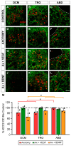
KCC2 immunofluorescence in the neuropil of extraocular motor nuclei 7 days after axotomy and axotomy with VEGF or BDNF treatment. Confocal images showing KCC2 immunofluorescence (green) in the oculomotor (OCM; (A,D,G,J)), trochlear (TRO; (B,E,H,K)), and abducens (ABD; (C,F,I,L)) nuclei. Extraocular motoneurons were identified by ChAT (red). KCC2 immunofluorescence is shown for control (AC), 7 days after axotomy (DF), and 7 days after axotomy (Ax) + VEGF (GI) or axotomy + BDNF (JL). The dashed white lines in (C,F,I,L) delimit the genu of the facial nerve. (M) Bar chart illustrating KCC2 optical densities (ODs) in the neuropil of the oculomotor, trochlear, and abducens nuclei 7 days after axotomy and axotomy with VEGF or BDNF delivery. Data are expressed as percentages relative (Re) to the control side (100%, dashed horizontal line). The following symbols were used to indicate significant differences between treatments within the same motor nucleus: *, significant difference (p < 0.05) compared to control; §, significant difference (p < 0.05) with respect to axotomy + VEGF; †, significant difference (p < 0.05) with respect to the axotomy + BDNF group. Hashtags were used to indicate significant differences (#, p < 0.05) between the three extraocular motor nuclei within the same treatment (two-way ANOVA test followed by Fisher´s post hoc test for multiple comparisons). Data correspond to mean ± SD. The number of animals per group was n = 3 (black dots). Scale bar is 75 µm in (L) for (AL).
Figure 3
Figure 3
KCC2 immunofluorescence in the somatic membrane of extraocular motoneurons 7 days after axotomy and axotomy plus VEGF or BDNF. (AL) Confocal images of oculomotor (OCM; (A,D,G,J)), trochlear (TRO; (B,E,H,K)), and abducens (ABD; (C,F,I,L)) motoneurons after immunostaining against KCC2 (green), in control (AC), 7 days after axotomy (DF), and 7 days after axotomy (Ax) + VEGF (GI) or axotomy + BDNF (JL). Inserts in each panel illustrate the same motoneuron/s identified by ChAT (red) immunoreactivity. Asterisks in (E,J,K) point to motoneurons whose KCC2 perisomatic staining was very faint. (M) Bar chart showing KCC2 optical density (OD) on the soma surface of extraocular motoneurons normalized with respect to (Re) the control side (100%, dashed horizontal line) and expressed as percentages in the following situations: axotomy, axotomy + VEGF, or axotomy + BDNF. To illustrate significant differences between treatments within the same motor nucleus, the following symbols were used: *, significant difference (p < 0.05) versus control; §, significant difference (p < 0.05) versus axotomy + VEGF; †, significant difference (p < 0.05) versus axotomy + BDNF. Hashtags were used to mark significant differences (#, p < 0.05) between the three types of extraocular motoneurons within the same treatment (two-way ANOVA test followed by Fisher´s post hoc test for multiple comparisons). Data correspond to mean ± SD. The number of animals per group was n = 3 (black dots). Scale bars are 5 µm in (L) for (AL) panels, and 5 µm in the insert in (L) for all inserts.
Figure 4
Figure 4
Effects of axotomy alone or together with the administration of VEGF or BDNF on the optical density of KCC2 in the neuropil of extraocular motor nuclei 15 days after axotomy. (AL) Confocal images showing KCC2 immunostaining (green) in the neuropil of the oculomotor (OCM; (A,D,G,J)), trochlear (TRO; (B,E,H,K)), and abducens (ABD; (C,F,I,L)) nuclei, in control (AC), and 15 days after axotomy (DF) or axotomy (Ax) + VEGF (GI) or BDNF (JL). ChAT immunofluorescence (red) was used to label the motoneurons. The dashed white lines in (C,F,I,L) delimit the genu of the facial nerve. (M) Bar chart of KCC2 optical density (OD) measurements in the neuropil of oculomotor, trochlear, and abducens nuclei 15 days after axotomy and after axotomy + VEGF or BDNF treatment. Data are represented as percentages relative (Re) to the control side (100%, dashed horizontal line). A two-way ANOVA test followed by Fisher’s post hoc test for multiple comparisons was used to detect significant differences between groups. The following symbols were used to indicate significant differences between treatments within the same motor nucleus: *, significant difference (p < 0.05) compared to control; §, significant difference (p < 0.05) with respect to axotomy + VEGF. Hashtags were used to indicate significant differences (#, p < 0.05) between the three extraocular motor nuclei within the same treatment. Data correspond to mean ± SD. The number of animals per group was n = 3 (black dots). Scale bar is 75 µm in (L) for (AL).
Figure 5
Figure 5
Analysis of the intensity of KCC2 immunofluorescence on the soma surface of extraocular motoneurons 15 days after axotomy and VEGF or BDNF administration. (AL) Confocal images of oculomotor (OCM; (A,D,G,J)), trochlear (TRO; (B,E,H,K)), and abducens (ABD; (C,F,I,L)) motoneurons (identified by ChAT immunostaining, in red, inserts in each panel) showing perisomatic immunofluorescence to KCC2 (green). Images correspond to control (AC) and 15 days after axotomy alone (DF) or axotomy (Ax) + VEGF (GI) or BDNF (JL) administration. Asterisks in (D,E,J,K,L) indicate motoneurons with very low levels of KCC2 immunostaining around their plasma membrane. White asterisks indicate cells with low perisomatic KCC2 labeling. (M) Bar chart showing KCC2 optical densities (ODs) on the soma surface of oculomotor, trochlear, and abducens motoneuron cell bodies. Data are expressed as percentages relative (Re) to the control side (100%, dashed horizontal line). A two-way ANOVA test followed by Fisher´s post hoc test for multiple comparisons was used to detect significant differences between groups. To indicate significant differences between experimental situations within the same motor nucleus, we used the following symbols: *, significant difference (p < 0.05) compared to the control; §, significant difference (p < 0.05) relative to the axotomy + VEGF situation; †, significant difference (p < 0.05) relative to the axotomy + BDNF situation. Hashtags illustrate significant differences (#, p < 0.05) between the three motoneuronal types within the same experimental situation. Data correspond to mean ± SD. The number of animals per group was n = 3 (black dots). Scale bars are 5 µm in (L) for (AL), and 5 µm in the insert in (L) for all inserts.
Figure 6
Figure 6
Schematic illustration of the present results at 15 days post-lesion (when KCC2 levels decreased to a minimum). (A) Control extraocular motoneurons (oculomotor, trochlear, and abducens) contained the cotransporter KCC2 in the plasma membrane of their dendrites (neuropil; in green) and cell bodies (in blue). These motoneurons receive neurotrophic factors retrogradely from their target muscle. (B) After axotomy, motoneurons were deprived of neurotrophic retrograde supply (red x). Muscles were also removed (dashed brown lines). Axotomy induced a remarkable decay in the levels of KCC2 in the neuropil and on soma surface of motoneurons. The only exception was the abducens motoneurons at the somatic surface level. (C) VEGF administration to axotomized motoneurons prevented the KCC2 decrease induced by lesion, at the level of the neuropil and soma surface. (D) By contrast, BDNF administration to axotomized motoneurons did not modify the injury-induced low KCC2 levels observed in the neuropil and on the soma surface. However, it should be emphasized that, in the particular case of the somatic membrane of abducens motoneurons, whose KCC2 levels did not change after injury, BDNF caused a significant downregulation of this cotransporter. Taken together, our results suggest that VEGF is an important neurotrophic factor involved in maintaining adequate levels of KCC2 along the somatodendritic membrane of these motoneurons, while BDNF might play a relevant role mediating the downregulation of KCC2 after injury.

Similar articles

References

    1. Akita T., Fukuda A. Intracellular Cl− dysregulation causing and caused by pathogenic neuronal activity. Pflug. Arch. 2020;472:977–987. doi: 10.1007/s00424-020-02375-4. - DOI - PubMed
    1. Virtanen M.A., Uvarov P., Mavrovic M., Poncer J.C., Kaila K. The multifaceted roles of KCC2 in cortical development. Trends Neurosci. 2021;44:378–392. doi: 10.1016/j.tins.2021.01.004. - DOI - PubMed
    1. Pressey J.C., de Saint-Rome M., Raveendran V.A., Woodin M.A. Chloride transporters controlling neuronal excitability. Physiol. Rev. 2023;103:1095–1135. doi: 10.1152/physrev.00025.2021. - DOI - PubMed
    1. Kim H.R., Martina M. Bidirectional Regulation of GABAA Reversal Potential in the Adult Brain: Physiological and Pathological Implications. Life. 2024;14:143. doi: 10.3390/life14010143. - DOI - PMC - PubMed
    1. Rivera C., Voipio J., Payne J.A., Ruusuvuori E., Lahtinen H., Lamsa K., Pirvola U., Saarma M., Kaila K. The K+/Cl− co-transporter KCC2 renders GABA hyperpolarizing during neuronal maturation. Nature. 1999;397:251–255. doi: 10.1038/16697. - DOI - PubMed

LinkOut - more resources