. 2024 Sep 15;25(18):9948.
doi: 10.3390/ijms25189948.

Photo-Crosslinked Pro-Angiogenic Hydrogel Dressing for Wound Healing

Affiliations

Photo-Crosslinked Pro-Angiogenic Hydrogel Dressing for Wound Healing

Wang Zhang et al. Int J Mol Sci. .

Abstract

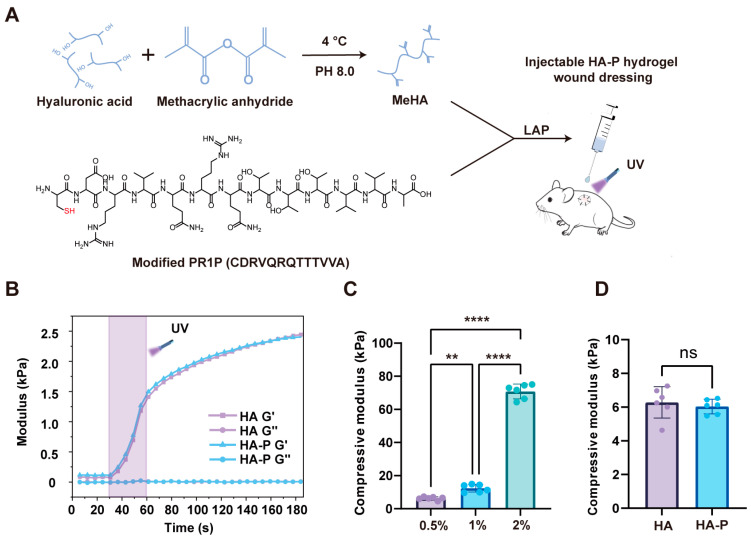
Severe burns are one of the most devastating injuries, in which sustained inflammation and ischemia often delay the healing process. Pro-angiogenic growth factors such as vascular endothelial growth factor (VEGF) have been widely studied for promoting wound healing. However, the short half-life and instability of VEGF limit its clinical applications. In this study, we develop a photo-crosslinked hydrogel wound dressing from methacrylate hyaluronic acid (MeHA) bonded with a pro-angiogenic prominin-1-binding peptide (PR1P). The materials were extruded in wound bed and in situ formed a wound dressing via exposure to short-time ultraviolet radiation. The study shows that the PR1P-bonded hydrogel significantly improves VEGF recruitment, tubular formation, and cell migration in vitro. Swelling, Scanning Electron Microscope, and mechanical tests indicate the peptide does not affect the overall mechanical and physical properties of the hydrogels. For in vivo studies, the PR1P-bonded hydrogel dressing enhances neovascularization and accelerates wound closure in both deep second-degree burn and full-thickness excisional wound models. The Western blot assay shows such benefits can be related to the activation of the VEGF-Akt signaling pathway. These results suggest this photo-crosslinked hydrogel dressing efficiently promotes VEGF recruitment and angiogenesis in skin regeneration, indicating its potential for clinical applications in wound healing.

Keywords: burn; hydrogel; methacrylate hyaluronic acid; prominin-1-binding peptide; vascular endothelial growth factor; wound healing.


Conflict of interest statement

The authors declare no conflicts of interest.

Figures

Figure 1
Figure 1
Fabrication of an injectable HA-P hydrogel wound dressing. (A) Schematic illustration of the synthesis of MeHA and in situ crosslinking with cysteine-modified PR1P to form a hydrogel wound dressing. (B) Real-time crosslinking rheological measurements of HA and HA-P hydrogels (0.5% w/v) with 30 s exposure to UV radiation. (C) Compressive modulus of HA hydrogels with different material concentrations (gelation with 30 s exposure to UV radiation). (D) Compressive modulus of HA and HA-P hydrogels (0.5% w/v, with 30 s exposure to UV radiation) (mean ± SD, n = 6, ** p < 0.01, **** p < 0.0001, ns, not statistically significant).
Figure 2
Figure 2
Characterization of HA and HA-P hydrogels. (A,B) SEM micrographs and quantification of the average pore size of freeze-dried HA hydrogels (0.5% w/v) with 30 s, 60 s, and 90 s of UV exposure. (C,D) SEM micrographs and quantification of the average pore size of HA and HA-P hydrogels (0.5% w/v, with 30 s of UV exposure). (E,F) Swelling ratios of HA hydrogels with various UV exposure times and material concentrations. (G) Swelling ratios of HA and HA-P hydrogels (0.5% w/v, with 30 s of UV exposure) (mean ± SD, n = 3, *** p < 0.001, **** p < 0.0001, ns, not statistically significant, scale bar, 100 μm).
Figure 3
Figure 3
VEGF recruitment and in vitro angiogenic effect of HA-P hydrogels. (A) Schematic illustration of VEGF recruitment assay. (B) Quantitative analysis of the maintained VEGF within hydrogels shows the HA-P hydrogel binds more VEGF than HA hydrogel does (n = 8). (C) Representative images of cell migration in a scratch wound healing assay after 0, 6, 12, and 24 h. (D) Quantitative analysis of the migration ratio shows HA-P hydrogel loaded with VEGF significantly promotes cell migration compared with the other groups. (E) Representative images of the tube formation of HUVECs. (F,G) Quantitative analysis of capillary length and the number of branch points of the tubule network. The capillary length and branch points in HA-P hydrogels are significantly higher than in the other groups (mean ± SD, n = 3, ** p < 0.01, *** p < 0.001, **** p < 0.0001, scale bar, 200 μm).
Figure 4
Figure 4
HA-P hydrogel dressing promotes wound regeneration in burns. (A) Representative photos exhibit the wound healing process. (B) Quantitative analysis of residual wound area (%) up to 14 days. HA-P hydrogel treatment shows significant acceleration of healing compared to the control group after day 6. (C) Representative images of H&E staining and (D) Masson’s trichrome staining of the wounds at 14 days post-wounding (scale bar, 500 μm). (E) Quantitative analysis of epithelium thickness and (F) collagen density indicates less epidermis hyperplasia and increased collagen deposition in the HA-P hydrogel treatment group (mean ± SD, n = 8–10, * p < 0.05, ** p < 0.01, ns, not statistically significant).
Figure 5
Figure 5
HA-P hydrogel dressing enhances angiogenesis and reduces myofibroblasts in burns. (A) Representative images of the CD31+ staining (green) of different groups at day 14 post-wounding. The nucleus was stained with DAPI (blue). (B,C) Stereological quantification of the surface area and length density of vasculature demonstrates a significant enhancement in angiogenesis for HA-P hydrogel compared with the control group. (D) Representative images of α-SMA+ staining (red) at day 14 post-wounding. The nucleus was stained with DAPI (blue). (E) Quantitative analysis of the positive area of α-SMA shows the HA-P hydrogel treatment significantly reduces myofibroblasts’ regeneration (mean ± SD, n = 8–10, ** p < 0.01, **** p < 0.0001, ns, not statistically significant, scale bar, 100 μm).
Figure 6
Figure 6
HA-P hydrogel dressing promoted angiogenesis via activation of the VEGF–Akt signaling pathway. (A) Schematic illustration of the molecular mechanism for HA-P hydrogel dressing which activates the VEGF–Akt signaling pathway in wound healing. (B) Representative images of Western blotting of Akt, p-Akt, and VEGFA in wounds at day 14 post-wounding. (C,D) Quantitative results of Western blotting show that the HA-P hydrogel treatment significantly increases the relative protein expression level of VEGFA and the relative expression ratio of p-Akt/Akt (mean ± SD, n = 8–10, * p < 0.05, ns, not statistically significant).
Figure 7
Figure 7
HA-P hydrogel dressing promotes wound healing in a full-thickness excisional wound model. (A) Representative images of the healing process up to 14 days post-wounding. (B) Wound closure curves of different groups show a significant acceleration of healing with the HA-P hydrogel treatment compared to the HA hydrogel and control group from day 4. (C) Representative images of CD31+ (green) and α-SMA+ (red) staining at day 14 post-wounding. The nucleus was stained with DAPI (blue). (D,E) Quantitative analysis indicates the HA-P hydrogel treatment significantly improves the angiogenesis and (F) reduces myofibroblasts’ regeneration (mean ± SD, n = 6, ** p < 0.01, **** p < 0.0001, ns, not statistically significant, scale bar, 100 μm).

Similar articles

Cited by

References

    1. Tottoli E.M., Dorati R., Genta I., Chiesa E., Pisani S., Conti B. Skin Wound Healing Process and New Emerging Technologies for Skin Wound Care and Regeneration. Pharmaceutics. 2020;12:735. doi: 10.3390/pharmaceutics12080735. - DOI - PMC - PubMed
    1. Sun B.K., Siprashvili Z., Khavari P.A. Advances in skin grafting and treatment of cutaneous wounds. Science. 2014;346:941–945. doi: 10.1126/science.1253836. - DOI - PubMed
    1. Greenhalgh D.G. Management of Burns. N. Engl. J. Med. 2019;380:2349–2359. doi: 10.1056/NEJMra1807442. - DOI - PubMed
    1. Radzikowska-Büchner E., Lopuszynska I., Flieger W., Tobiasz M., Maciejewski R., Flieger J. An Overview of Recent Developments in the Management of Burn Injuries. Int. J. Mol. Sci. 2023;24:16357. doi: 10.3390/ijms242216357. - DOI - PMC - PubMed
    1. Zwierello W., Piorun K., Skorka-Majewicz M., Maruszewska A., Antoniewski J., Gutowska I. Burns: Classification, Pathophysiology, and Treatment: A Review. Int. J. Mol. Sci. 2023;24:3749. doi: 10.3390/ijms24043749. - DOI - PMC - PubMed

MeSH terms

LinkOut - more resources